
Advantages and Disadvantages of Treatment Options for Congenitally Missing Lateral Incisors
Thomas E. Dudney, DMD
The treatment of congenitally missing maxillary lateral incisors presents unique challenges to the dental profession and often requires a coordinated interdisciplinary approach involving orthodontists, oral surgeons, periodontists, and restorative dentists to achieve optimal esthetic and functional results. Careful diagnosis and information gathering as well as communication with the patient— and often, depending on age, their parents—is necessary to formulate a treatment plan that will accomplish the desired results and satisfy patient expectations. There are many factors that affect treatment options, such as occlusion; alignment of teeth; patient age; available space; alveolar bone thickness; facial profile; shape, color, and size of the canines; lip position; gingival display; and condition of adjacent teeth.1-4 An ideal treatment plan should take into account all of these factors to achieve the esthetic and functional goals with the least-invasive option available.5 This article will not recommend a particular treatment modality, but identify the available options and discuss their advantages and disadvantages.
Space Closure with Canine Substitution
The two main treatment options include either space opening with prosthetic replacement or space closure with canine substitution.1,4 In ideal situations where patients have a balanced or slightly convex facial profile; an angle class II malocclusion with no mandibular crowding or an angle class I malocclusion with mandibular crowding necessitating extractions; and small canines that are lighter in color, space closure may be a viable alternative.6 The advantages of canine substitution are that natural teeth are biocompatible and preferable long term and the finished result is permanent, negating the need for future prosthetic replacement.1,2 Furthermore, even though canine-protected occlusion is not possible, long-term studies have shown that space closure using a premolar is equally sound occlusally and preferable periodontally to space opening. The disadvantages of canine substitution are the need for certain conditions to exist as previously stated, adverse effects on dentoalveolar and facial esthetics as a result of the canine prominence being moved mesially, the tendency for space to sometimes reopen, and very large or dark canines that are difficult to reshape or bleach to an acceptable shade and may require a restoration (ie, porcelain veneer) to achieve the desired esthetic result (Figure 1A and Figure 1B).
Space Opening with Prosthetic Replacement
Historically, options for space opening with prosthetic replacement were limited to removable partial dentures and conventional fixed bridges. Neither option was very appealing and in the past made space closure with canine substitution a more attractive alternative. The only advantage of removable partial dentures was that they were far more conservative than fixed bridges. However, their disadvantages included their bulky and cumbersome design, which made it difficult to achieve esthetic results, and the fact that patients disliked wearing them.
Although conventional fixed bridges were more comfortable and esthetic than removable partial dentures, they required gross reduction of healthy tooth structure, which is generally contraindicated in young patients. With today’s materials, fixed bridges offer excellent results from esthetic and functional perspectives, but because they still require an amount of tooth reduction their use should be limited to situations where a conventional bridge already exists or the condition of the abutment teeth (ie, wear, caries, fracture, etc) would dictate a more aggressive preparation.
The first attempt at a conservative fixed restorative option was the Maryland bridge, which had metal wings that bonded to the lingual surface of the central and canine. The advantage of a Maryland bridge was that it was minimally invasive, but the disadvantages included compromised esthetics from the metal wings showing through translucent enamel and the inability of the rigid metal wings to flex with the slight mobility of the anterior teeth, often leading to debonding.7 In recent years two more options for prosthetic replacement have gained favor. One is the fiber-reinforced resin-bonded bridge and the other is the single-tooth implant. Both options offer a conservative approach with acceptable esthetics and, although each option has certain advantages, each also has disadvantages.
The Fiber-Reinforced Resin-Bonded Bridge
The fiber-reinforced resin-bonded bridge, sometimes referred to as the Encore bridge,7 is based on the same principle as the Maryland bridge. Instead of metal wings, this type of bridge incorporates laboratory-processed composite resin with fiber reinforcement in the form of a lingual framework with a ceramic veneer bonded to the facial of the pontic. This design ensures long-term esthetics and vitality of the restoration.7 Unlike metal, the fiber-reinforced resin framework readily bonds to enamel and dentin and has the strength and elasticity to resist fracture and debonding, even in the presence of slight mobility of the anterior teeth.
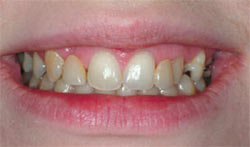
Furthermore, because the material is tooth-colored, the problem of discoloration and graying of enamel caused by metal show-through is eliminated (Figure 2A and Figure 2B). In cases where patients desire a more comprehensive smile enhancement procedure in addition to replacing the congenitally missing lateral incisors, this technique works well because porcelain veneers can be placed on the natural teeth as well as the pontics of the resin frameworks, thus giving the restorative team control of the size, shape, position, and color of the restorations in the final smile (Figure 3A; Figure 3B; Figure 3C).
Fiber-reinforced resin-bonded bridges with ceramic overlays on the pontics can be an excellent treatment option, but there are some disadvantages of this procedure as well. While they are much less invasive than conventional three-unit or even two-unit canine cantilevered fixed bridges, these bridges do require some tooth preparation on the lingual and interproximal surfaces of the abutment teeth. Also, because of their high failure rate when subjected to excessive lateral forces, these bridges are not recommended for pa- tients with a deep overbite relationship.5 Although different preparation designs exist, the one that incorporates as much of the lingual surface as possible (a depth of about 1 mm) and has a horizontal central groove for strength and a proximal box design for increased thickness and fiber concentration in the connector sites seems to achieve the best long-term results,7 but also requires the most preparation of healthy tooth structure. Furthermore, this type of fixed restorative option likely would have to be remade several times over the lifetime of a young patient, and the possibility of connector site fractures, material delamination, or debonding still exists. However, in cases where canine substitution or single-tooth implant replacement are contraindicated or the patient chooses not to go through the necessary protocol for implant placement, the fiber-reinforced resin-bonded bridge may be the best restorative option.7
The Single-Tooth Implant
A more recent option for treating congenitally missing lateral incisors, and one that currently is recommended often, is the single-tooth implant. Over the past several years, the predictability and long-term success rates of implants have made them an obvious restorative choice,5 especially when teeth adjacent to the space are healthy, of normal size and shape, and unrestored.8,9 Furthermore, placement of an implant may provide a functional stimulus to help preserve bone and prevent resorption.10 However, when choosing the single-tooth implant as a restorative op- tion, several factors must be taken into ac- count such as growth considerations, space requirements, and site development.3
Because an implant acts essentially like an ankylosed tooth, any vertical alveolar growth and eruption of teeth would cause a discrepancy between the gingival margin of the natural tooth and the implant. Therefore, implant placement should occur only after growth has been completed,2-4 and it has been suggested that neither chronological age nor hand-wrist radiographs are reliable enough to make that determination.3 Instead it would be best to compare superimposed cephalometric radiographs taken at 1-year intervals until no growth changes are detected.3,9 Also, the amount of space between the roots is critical to successful implant placement, and orthodontic intervention usually is necessary to achieve not only the amount of interradicular space needed, but also the proper root angulation.2,3 Because orthodontic treatment usually occurs at an early age, several years of maintenance therapy may be required until the appropriate age for implant placement. It is also important to maintain proper spacing for ideal tooth proportions of the final restoration.
In addition to the tooth width requirements for mesiodistal spacing, the alveolar width in a buccolingual direction must be adequate for implant placement. Often an additional surgical appointment is necessary to graft or augment the alveolar ridge before an implant can be placed. It has been suggested in the literature that by allowing or guiding the eruption of the canines into the lateral position and orthodontically moving them to their natural position, the necessary amount of buccolingual alveolar thickness for implant placement can be achieved naturally, without the need to perform any ridge augmentation.2,4,7 Although not completely understood, it has been shown that very little, if any, resorptive change in alveolar bone width is observed when space is opened orthodontically compared with the decrease in alveolar ridge width after extraction of maxillary anterior teeth. However, a disadvantage of orthodontic canine distalization for implant site development is the potential for loss of arch length when the canines are allowed to erupt mesially.3
While single-tooth implants as a prosthetic replacement for congenitally missing lateral incisors can conservatively satisfy the esthetic, functional, and biologic goals of treatment, they are not without disadvantages and often require very careful case selection and a diligent and well-coordinated interdisciplinary approach. In addition to site development, space requirements, and age restrictions because of the necessity for growth completion as conditions that must be met for successful treatment, implants as a restorative option have the potential for esthetic failure as a result of gingival darkening, exposure at the margin from gingival recession, incisal edge and gingival discrepancies from long-term facial growth and vertical movement of teeth,11 and the difficulty of matching natural tooth color and translucency with an implant crown. When restoring congenitally missing lateral incisors with implants, or any type of restorative option, the patient should be committed to a lifetime of wearing a prosthesis in the area of the mouth where esthetics are critical and not always easy to control.11
Conclusion
The treatment of congenitally missing maxillary lateral incisors is challenging and complex, requiring very careful treatment planning, communication with the patient, and often the coordinated interdisciplinary efforts of the orthodontist, periodontist, surgeon, and restorative dentist. The two categories of treatment options include space closure with canine substitution and space opening with prosthetic replacement. At the present time, the two most recommended procedures for prosthetic replacement are the single-tooth implant and the fiber-reinforced resin-bonded bridge with a ceramic overlay. Although this article did not recommend a particular treatment modality or extol the virtues of one choice over another, it identified the available options and discussed the advantages and disadvantages of each one. There is probably not one treatment choice for this particular clinical situation that is clearly the best option for each individual case. It is only after weighing all of the options available, analyzing the conditions that exist, and consulting with the patient and other specialists that a method of treatment should be chosen.
References
1. Sabri R. Management of missing maxillary lateral incisors. J Am Dent Assoc. 1999;130 (1):80-84.2. Spear FM, Mathews DM, Kokich VG. Interdisciplinary management of single tooth implants. Semin Orthod. 1997;3(1): 45-72.
3. Chan E, Darendeliler MA, Vickers D, et al. Implants and orthodontics. Brighter Futures. Newsletter of the Australian Society of Orthodontists. 2006;3:1-4.
4. Kokich VO Jr. Early management of congenitally missing teeth. Semin Orthod. 2005; 11(3):146-151.
5. Kinzer GA, Kokich VO Jr. Managing congenitally missing lateral incisors. Part II: tooth-supported restorations. J Esthet Restor Dent. 2005;17(2): 76-84.
6. Kinzer GA, Kokich VO. Managing congenitally missing lateral incisors. Part 1: canine substitution. J Esthet Restor Dent. 2005; 17(1):5-10.
7. Hornbrook D. The fiber-reinforced resin-bonded bridge. Dental Products Report. February 2004. Available at: www.dentalproducts.net/xml/display.asp?file=2247&bhcp=1. Accessed June 26, 2008.
8. The biologic basis of orthodontic therapy. In: Proffit WR. Contemporary Orthodontics. 3rd ed. St. Louis, MO: CV Mosby; 2000: 616.
9. Kokich VG. Maxillary lateral incisor implants: planning with the aid of orthodontics. J Oral Maxillofac Surg. 2004;62(9 Suppl 2):48-56.
10. Balshi TJ, Wolfinger GJ. Single tooth replacement: Fixed partial denture vs single tooth implants. Valley Forge Dental Journal. Spring 1997. Available at: https://www.dentalimplants-usa.com/Treatment/Implants/single.html. Accessed June 26. 2008.
11. Zachrisson BU, Stenvik A. Single implants—optimal therapy for missing lateral incisors? Am J Orthod Dentofacial Orthop. 2004;126 (6):A13-A15.
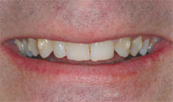 | 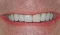 | |
| Figure 1A Unilateral congenitally missing lateral incisor with canine substitution (tooth No. 11 moved into site No. 10). | Figure 1B The canine was reshaped and the smile was restored with porcelain veneers. | |
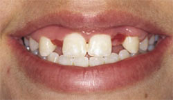 | 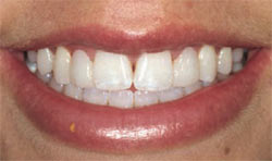 | |
| Figure 2A Bilateral congenitally missing lateral incisors. | Figure 2B Fiber-reinforced resin-bonded bridges with ceramic veneers on the pontics at site Nos. 7 and 10. | |
 | 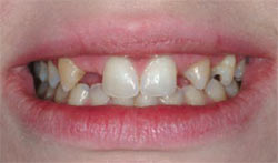 | |
| Figure 3A Bilateral congenitally missing lateral incisors with a removable partial denture in place. | Figure 3B Facial view with the removable partial denture out of the mouth. | |
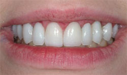 | ||
| Figure 3C Fiber-reinforced resin-bonded bridges and ceramic veneers on the pontics (site Nos. 7 and 10) and natural teeth restored with veneers (site Nos. 4, 5, 6, 8, 9, 11, 12, and 13). | ||
| About the Author | ||
 Thomas E. Dudney, DMD Thomas E. Dudney, DMD Private Practice Aesthetic, General, and Restorative Dentistry Alabaster, Alabama | ||

