You must be signed in to read the rest of this article.
Registration on CDEWorld is free. You may also login to CDEWorld with your DentalAegis.com account.
Determining the type of restoration that should be placed in implant dentistry is a decision that is based on many factors, including but not limited to the occlusal clearance, esthetics, anatomy, and surgical placement of the implant or implants. For each case, clinicians are forced to choose between cement-retained and screw-retained restorations. This decision is often a difficult one, particularly in the anterior maxilla, which poses a number of challenges anatomically and prosthetically. Cement-retained implant restorations are cited for their ability to maintain the anatomy because they do not need a screw access, for their familiar cementation protocol, for their ability to correct angulation errors, and most notably, for their esthetics. However, retaining implant-supported restorations with cement has also been documented as a major drawback because it severely hinders retrievability of the prosthetic screw and excess cement has been noted in the literature as a frequent cause of peri-implantitis.1
Critical factors in the survivability of implants include tension distribution, bone quality, and bone quantity. Therefore, implants are surgically placed where the available bone permits, but this may result in a malpositioning that can leave the restoring dentist with limited options. Screw-retained restorations, which are often noted for their improved retrievability, were traditionally difficult to place in the anterior maxilla due to the precise placement required by straight screw channels.2 As clinicians sought to merge the esthetics of cement-retained restorations with the practicality and predictability of screw-retained ones, the angulated screw channel was developed as a viable option to avoid unsightly screw access openings on the incisal edges or facial surfaces of anterior restorations.
Screw Channel Angulation
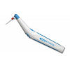
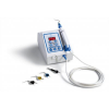
The concept of the angulated screw channel was first introduced in 2004 with an abutment that permitted a deviation of up to 28° for screw-retained restorations. Following its release, other manufacturers developed similar options, and currently, angulations of up to 30° can be corrected with angulated screw channels, depending on the manufacturer of the implant components. Restorations fabricated in this manner cannot be placed with a traditional driver. They require an off-axis driver that is able to engage the prosthetic screw in order to achieve final torque values (Figure 1).3 Although the body of literature regarding this paradigm is growing, thus far, it is favorably in support of its use. Placing screw-retained implant restorations with angulated screw channels comes with several key advantages. First, the elimination of cement facilitates a better long-term prognosis for implant-retained restorations. In addition, by avoiding the need to over-contour the restoration, a more hygienic one can be delivered that will have a favorable outcome on the surrounding soft tissue. The esthetics are also improved because the screw access of a screw-retained crown may now be placed in a less visible location. This is important because observational studies using cone-beam computed tomography (CBCT) analysis have shown that although screw-retained restorations (straight channel or angulated channel) may be achievable in approximately 75% of cases, they are only used in approximately 25% of cases, and cement-retained ones may have compromised results.4 The current literature supporting the use of screw-retained implant restorations suggests that as clinicians become more familiar with this more modern approach and technique, more predictable esthetic and peri-implant health outcomes may be achieved.5 Restorations with angulated screw channels can be used anteriorly or posteriorly in either arch.
Materials Science and Testing
Because there is adequate research demonstrating that implants best transmit occlusal loads along the long axes of their fixtures, there were originally concerns regarding the long-term survival of restorations with angulated screw channels.6 However, angled abutments, which predated the angulated screw channel and ultimately led to its development, have had their viability evaluated in materials tests. Finite element analysis has been used to evaluate the effects that angled abutments have on the prosthetic components of the implant and the surrounding bone. Finite element analysis uses a virtual environment created by a computer to simulate and predict how an object will respond to applied stress in the real world. In one study utilizing this analysis that showed that increasing implant diameter width was associated with as much as a 3.5-fold reduction in crestal strain and that increasing implant length was associated with as much as a 1.65-fold reduction in crestal strain, when the effects of oblique forces on cortical and cancellous bone were compared with the effects of those along the long axis, they were found to be within physiological limits as long as there was adequate bone.7 Furthermore, in a similar study comparing screw-retained versus cement-retained zirconia restorations, the screw-retained restorations demonstrated less stress transfer to the surrounding environment, which supports their use.8
Photoelastic stress analysis is another common form of testing in which the object to be tested is placed in or coated with a transparent epoxy. This material exhibits an optical characteristic called birefringence or double refraction, which essentially splits a ray of light into two different beams, when it is placed under stress. A common polymer familiar to dentists that displays this phenomenon is polycarbonate. In photoelastic stress analysis, plane-polarized light is passed through the system while it is stressed, and a colored set of patterns called isochromatic fringes is formed that provides a visual representation of the stress. In general, frequent and closely spaced fringes indicate areas of high stress, whereas black areas can sometimes indicate low stress.9-11 Although these patterns require careful interpretation, they can provide useful data for materials testing. A strain is defined as the amount of deformation per unit length of an object when load is applied. In one study that used photoelastic stress analysis to measure the strain placed by implant abutments on surrounding bone in micro-strains (10-6), the researchers found that when compared with nonangulated restorations, angulated restorations placed an increased strain on the bone; however, there was no statistically significant difference in the time to failure, and both restoration types generally existed within physiologic limits. Because bone density may play a role in survival when comparing nonangulated restorations with angulated restorations due to the difference in the stresses, the researchers recommended further study to evaluate the effects of immediate loading.12,13 Another study involving photoelastic stress analysis demonstrated increased strain when forces were applied at a 30° angle,11 and others have confirmed this finding at less extreme angulations, including one study that demonstrated that an angulation of 25° resulted in an approximate 4.5-fold increase in strain whereas an angulation of 15° resulted in an approximate 3.0-fold increase in strain.14 However, both studies reported that the survival rates for restorations with angulated screw channels were similar to those of restorations with straight screw channels, so they should be indicated for use.
Although angulated restorations may place an increased load prosthetically and biologically, their survival rates in the current literature appear to be consistent with the conventional paradigm. Restorations with angulated screw channels also show comparable fracture resistance to those with straight screw channels, with complications that are common among different implant connection types, such as screw loosening, being reported as a result of a variety of clinical factors, including preload, angle, and screw design.15 Further research is needed to make clinical recommendations regarding immediate loading, pontic length, and other clinical scenarios that may result in increased strain.
Angulated Channel Zirconia Restorations
Zirconia restorations, particularly full contour ones, have become common clinically and are often desired for their combination of esthetics, fracture resistance, flexural strength, and resistance to chipping.16 For dental purposes, zirconia refers to zirconium dioxide (ZrO2), which sequentially transitions through three phases as it is brought to room temperature: cubic, tetragonal, and monoclinic. The monoclinic phase exists between approximately 1,170°C and room temperature. When zirconia is cooled from the tetragonal phase to the monoclinic phase, a volume expansion of approximately 4% occurs, which is called martensitic transformation.17 This is problematic because unwanted imperfections and cracks may propagate throughout the material. Therefore, yttrium oxide is incorporated into dental zirconia as a stabilizing agent so that the tetragonal phase can exist at room temperature. Second generation zirconia, or 3 mol% yttria-stabilized zirconia (3Y-TZP), is noted for its strength, but because clinicians sought greater translucency for uses in the anterior, a third generation, or 5 mol% yttria-stabilized zirconia (5Y-TZP) was developed for more lifelike characteristics. The increase in yttria results in increased translucency because it allows for a greater percentage of the cubic phase to be present, which has greater volume in size and does not scatter light as much, so more of it passes through.18 Unfortunately, when the yttria content of zirconia is increased, there is a noted decrease in the strength of the material.
Because the use of zirconia is increasing in popularity for restorations of all kinds, it is a consideration for implant restorations with angulated screw channels. In a 3-year prospective cohort study that measured the effect of monolithic zirconia restorations with angulated screw channels on crestal bone loss, the restorations exhibited a survival rate of 96%, and two incidences of screw loosening were the only mechanical complications reported.19 Furthermore, in a 42-month retrospective study of zirconia restorations with angulated screw channels that reported an overall survival rate of 92%, monolithic crowns demonstrated a higher survival rate (95%) when compared with those that were veneered with porcelain (90%). Restorations placed in both the anterior and posterior regions showed high survival rates.20 Posterior angulated screw channel restorations have been shown to be useful because they provide greater and more convenient access for both clinical placement and future retrievability. A study that evaluated full-contour zirconia crowns with angulated screw channels placed in the posterior maxilla and mandible at 1-year follow-up found that although there was an average of 0.16 mm of marginal bone loss, there were no failures. The patients selected for the study had implant sites that were free from infection and demonstrated adequate bone height.21
Case Report 1
A 68-year-old woman presented with decay affecting teeth Nos. 5 and 6 to the extent that they were non-restorable. Both teeth were extracted, and the sites were grafted. After a healing period, an implant was placed into each site, and custom contoured healing abutments were delivered (Figure 2). After another healing period, the emergence profile established would ensure that there was ideal contour of the restorations, passive fit to minimize or eliminate tissue blanching on delivery, and because a digital workflow was to be used, ideal visualization of the scan bodies (Figure 3). Appropriate scan bodies were hand tightened into the fixtures and upper and lower full-arch scans were acquired (Figure 4). During digital design, it was noted that although the tooth No. 5 site could accommodate a straight-channel screw-retained restoration, using the same design for tooth No. 6 would place the access on the incisal edge (Figure 5). This would be an esthetically challenging location as well as subject the access to a higher rate of wear during lateral excursive movements. Therefore, a design with an angulated screw channel was chosen for tooth No. 6, which addressed the complications that would occur with a straight channel (Figure 6). Although both implants were the same platform size and from the same manufacturer, due to the nature of attaching the restorations, on delivery, tooth No. 5 was torqued to 35 Ncm while tooth No. 6 was torqued to 25 Ncm (Figure 7). The screw channels were then blocked with polytetrafluoroethylene (PTFE) tape, and the access holes were restored with a universal composite. The end result blended with the surrounding dentition in form, function, and esthetics (Figure 8).
Case Report 2
A 55-year-old woman presented to the practice missing tooth No. 10, which had previously fractured due to decay. In addition, teeth Nos. 7 through 9 had large multi-surface fillings that were failing (Figure 9). Due to the esthetic demand and the anticipated amount of missing tooth structure, a diagnostic wax-up was digitally designed (Figure 10). The diagnostic wax-up was then 3D printed and used to fabricate a clear stent. During the preparation phase, the stent was intermittently used to verify that the amount of reduction was adequate and in accordance with the digital design (Figure 11). Temporary crowns were fabricated from a bis-acryl material and delivered, and the patient was dismissed to be seen 2 weeks later to ensure that there were no changes in the soft-tissue architecture. A bone-level implant was placed in the tooth No. 10 site. Due to the depth of the fixture, a conventional impression coping would provide a more ideal fit and visualization of site No. 10 for the design of the final implant-supported restoration. Therefore, a final upper impression was taken using a polyether impression material. During design, an angulated screw channel was chosen to place the access opening in its ideal position (Figure 12). Because the final restorations for teeth Nos. 7 through 9 were going to be conventionally cemented, the order of delivery was planned such that the tooth No. 10 restoration would be delivered last. This allowed for the complete removal of any excess cement from teeth Nos. 7 through 9 so that it would not compromise the implant site (Figure 13). The patient's smile exhibited an ideal amount of display, and the restorations blended well with the surrounding dentition (Figure 14).
Conclusion
Angulated screw channels allow for screw-retained restorations to be placed in cases in which cement-retained ones may traditionally have been used, which may improve patients' long-term prognoses. It is important to be knowledgeable regarding the armamentarium that is needed to place these restorations as well as the appropriate torque values, which may differ from those of their straight-channel counterparts. The current research is overall supportive of the use of screw-retained implant restorations with angulated screw channels in a variety of clinical scenarios. However, the growing body of literature will help to establish evidence-based guidelines regarding immediate loading, bone density, and restorative designs, preserving the advantages that come with screw-retained crown and bridge dentistry.
Queries regarding this course may be submitted to authorqueries@broadcastmed.com
About the Author
Stephen Dadaian, DDS
Private Practice
Cresskill, New Jersey
References
1. Rokaya D, Srimaneepong V, Wisitrasameewon W, et al. Peri-implantitis update: risk indicators, diagnosis, and treatment. Eur J Dent. 2020;14(4):672-682.
2. Safi Y, Amid R, Zadbin F, et al. The occurrence of dental implant malpositioning and related factors: a cross-sectional cone-beam computed tomography survey. Imaging Sci Dent. 2021;51(3):251-260.
3. Berroeta E, Zabalegui I, Donovan T, Chee W. Dynamic abutment: a method of redirecting screw access for implant-supported restorations: technical details and a clinical report. J Prosthet Dent. 2015;113(6):516-519.
4. Edmondson EK, Trejo PM, Soldatos N, Weltman RL. The ability to screw-retain single implant-supported restorations in the anterior maxilla: a CBCT analysis. J Prosthet Dent. 2022;128(3):443-449.
5. Nastri L, Nucci L, Grassia V, Miraldi R. Aesthetic outcomes and peri-implant health of angled screw retained implant restorations compared with cement retained crowns: medium term follow-up. J Funct Biomater. 2021;12(2):35.
6. Song JY. Implant complications in bruxism patients. J Korean Assoc Oral Maxillofac Surg. 2021;47(2):149-150.
7. Petrie CS, Williams JL. Comparative evaluation of implant designs: influence of diameter, length, and taper on strains in the alveolar crest. A three-dimensional finite-element analysis. Clin Oral Implants Res. 2005;16(4):486-494.
8. Lee JH, Jang HY, Lee SY. Finite element analysis of dental implants with zirconia crown restorations: conventional cement-retained vs. cementless screw-retained. Materials (Basel). 2021;14(10):2666.
9. Hearn EJ. Mechanics of Materials 2. 3rd ed. Butterworth-Heinemann; 1997.
10. Marín-Miranda M, Wintergerst AM, Moreno-Vargas YA, er al. Photoelasticity for stress concentration analysis in dentistry and medicine. Materials (Basel). 2022;15(19):6819.
11. Ueda C, Markarian RA, Sendyk CL, Laganá DC. Photoelastic analysis of stress distribution on parallel and angled implants after installation of fixed prostheses. Braz Oral Res. 2004;18(1):45-52.
12. Clelland NL, Gilat A. The effect of abutment angulation on stress transfer for an implant. J Prosthodont. 1992;1(1):24-28.
13. Zanatta LC, Dib LL, Gehrke SA. Photoelastic stress analysis surrounding different implant designs under simulated static loading. J Craniofac Surg. 2014;25(3):1068-1071.
14. Brosh T, Pilo R, Sudai D. The influence of abutment angulation on strains and stresses along the implant/bone interface: comparison between two experimental techniques. J Prosthet Dent. 1998;79(3):328-334.
15. Rasaie V, Abduo J, Falahchai M. Clinical and laboratory outcomes of angled screw channel implant prostheses: a systematic review. Eur J Dent. 2022;16(3):488-499.
16. Borrero-Lopez O, Guiberteau F, Zhang Y, Lawn BR. Wear of ceramic-based dental materials. J Mech Behav Biomed Mater. 2019;92:144-151.
17. Resnik R. Misch's Contemporary Implant Dentistry. 4th ed. Mosby; 2020.
18. Stawarczyk B, Keul C, Eichberger M, et al. Three generations of zirconia: from veneered to monolithic. Part I. Quintessence Int. 2017;48(5):369-380.
19. Di Fiore A, Granata S, Monaco C, et al. Clinical performance of posterior monolithic zirconia implant-supported fixed dental prostheses with angulated screw channels: a 3-year prospective cohort study [published online ahead of print August 2, 2021]. J Prosthet Dent. 2021;doi: 10.1016/j.prosdent.2021.06.043.
20. Rella E, De Angelis P, Damis G, et al. The application of angulated screw-channels in metal-free, implant-supported restorations: a retrospective survival analysis. Materials (Basel). 2021;14(22):7006.
21. Pol CWP, Raghoebar GM, Maragkou Z, et al. Full-zirconia single-tooth molar implant-supported restorations with angulated screw channel abutments: a 1-year prospective case series study. Clin Implant Dent Relat Res. 2020;22(1):138-144.