
Immediate Loading of the Edentulous Mandible Using a Definitive Hybrid Prosthesis
R. Bruce Miller; Samuel Zfaz; Auchim Michael Renner
Although the oldest of all of the osseointegrated dental implant prostheses, the mandibular fixed-detachable hybrid prosthesis continues to evolve and improve, primarily indicated for edentulous or nearly edentulous patients with available bone anterior to the metal foramen and insufficient bone posterior.1 With rapid advances in the area of immediate loading of dental implants, the hybrid prosthesis has regained popularity as a predictable and less time-consuming treatment modality.
With success rates of immediately loaded implants in the anterior mandible equaling those of conventional-loading protocols,2,3 the clinical advantages of these techniques become apparent. For the nearly edentulous patient, the prospect of not having to wear an immediate interim mandibular denture during a 2- to 4-month integration period is a distinct advantage. In addition, the patient is able to function immediately with a fixed prosthesis. It is further advantageous for the patient if the definitive prosthesis can be immediately delivered, thereby eliminating the provisional stage and subsequent multiple postintegration appointments required to fabricate the definitive teeth.
If immediate loading of dental implants is going to be successful, careful presurgical prosthetic planning is essential. A critical determinant to the success of an immediately loaded prosthesis includes primary stability of the implants, which may be of concern when placing implants in extraction sockets. In addition, idealizing the patient’s occlusal scheme, vertical dimension, and tooth position should be performed before any surgical procedures.
Case Presentation
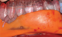
A 62-year-old woman presented with a chief complaint of dissatisfaction with her appearance and inability to chew (Figure 1). The patient was diagnosed with generalized moderate-to-severe periodontal disease, caries, and occlusal dysfunction (Figure 2). Her medical history was unremarkable. Several techniques for the immediate restoration of the edentulous mandible have been reported in the literature.2,3 The technique presented here describes the delivery of the definite fixed prosthesis within the 72-hour window for predictable osseointegration.
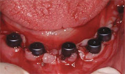
The patient’s remaining maxillary teeth (except tooth No. 11) were extracted and a maxillary immediate complete denture was delivered to idealize the occlusal plane and maxillary tooth position (Figure 3). A final impression was made of the mandibular arch, the cast was articulated on a semi-adjustable articulator, and a denture tooth set-up was completed (Figure 4). The set-up was duplicated in clear orthodontic resin for use as a surgical template and a custom tray for the final impression of the implants (Figure 5). The surgical procedure involved extraction of the remaining mandibular teeth and an alveolectomy to gain interarch space for the definitive prosthesis (Figure 6).4 Placement of five endosseous implants (Thommen Medical USA, Bartlesville, OK) anterior to the metal foramen was performed maximizing the anterior-posterior spread (Figure 7). An impression/centric relation record was made of the fixtures using the surgical template at the desired vertical dimension of occlusion (Figure 8 and Figure 9). Healing abutments were placed and the tissue was sutured using 4-0 silk sutures (Figure 10).
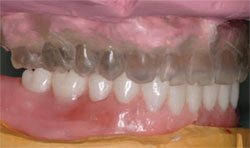
In the laboratory, the final impression was attached to the altered, mounted master casts and poured using the implant laboratory analogs (Thommen Medical). The framework was waxed according to the denture tooth index and cast in a high palladium alloy (Will-Ceram W-1, Ivoclar Vivadent, Amherst, NY) (Figure 11). The patient was recalled at 48 hours for try-in of the casting and wax tooth set-up. Passive seating of the framework was confirmed as well as verification of tooth position and centric relation. The prosthesis was removed and returned to the laboratory for processing using a pour technique (Palapress Vario®, Heraeus Kulzer, Inc, Armonk, NY).
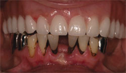
The prosthesis was delivered to the patient with postoperative instructions to include a soft diet (Figure 12). The prosthesis was left in place for 12 weeks for integration of the implants (Figure 13). All implants integrated without complications and the patient’s function was very good. This technique provided the patient with stable, functioning definitive teeth within 48 hours of the surgical procedure with an excellent prognosis.
CONCLUSION
In its most literal interpretation, immediate loading is defined as the placement of the restoration in function on the implants at the same clinical visit. Early loading implies functional loading no earlier than 3 weeks and up to 6 to 8 weeks postsurgery. Conventional loading allows for more traditional healing times of 3 to 6 months before loading. Delayed loading is used when extended healing periods (> 3 months) are required because of poor quality, the quality of the bone, and lack of stability of the implants. Today, numerous protocols exist for immediate loading of dental implants. The common goal is to provide to the patient a carefully planned functional and esthetic restoration that offers scientifically determined, predictable prognosis.
DISCLOSURE
The author receives a grant and research support from Thommen Medical.
REFERENCES
1. Adell R, Lekholm U, Rockler B, et al. A 15-year study of osseointegrated implants in the treatment of the edentulous jaw. Int J Oral Surg. 1981;10(6):387-416.
2. Cooper LF, Rahman A, Moriarty J, et al. Immediate mandibular rehabilitation with endosseous implants: simultaneous extraction, implant placement and loading. Int J Oral Maxillofac Implants. 2002;17(4):517-525.
3. Ganeles J, Rosenberg MM, Holt RL, et al. Immediate loading of implants with fixed restorations in the completely edentulous mandible: report of 27 patients from a private practice. Int J Oral Maxillofac Implants. 2001;16(3): 418-426.
4. AbuJamra NF, Stavridakis MM, Miller RB. Evaluation of interarch space for implant restorations in edentulous patients: a laboratory technique. J Prosthdont. 2000; 9(2):102-105.
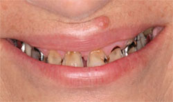 | 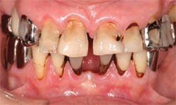 | ||||
| Figure 1 Patient’s pretreatment smile. | Figure 2 Pretreatment clinical presentation. | ||||
 |  | ||||
| Figure 3 Maxillary immediate complete denture. | Figure 4 Articulated casts with mandibular set-up. | ||||
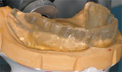 | 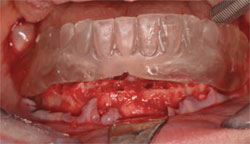 | ||||
| Figure 5 Surgical template/custom tray. | Figure 6 Surgical template indicating alveolar reduction for desired prosthetic interocclusal space. | ||||
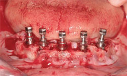 | 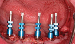 | ||||
| Figure 7 Implant placement maximizing anterior/posterior spread. | Figure 8 Open tray implant impression copings in place. | ||||
 |  | ||||
| Figure 9 Impression and centric relation recorded at the desired vertical dimension of occlusion. | Figure 10 Healing abutments in place. | ||||
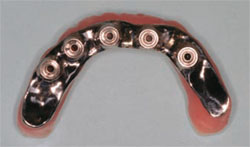 | 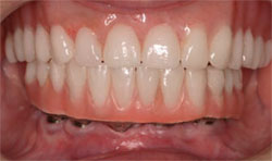 | ||||
| Figure 11 Completed cast substructure. | Figure 12 Clinical view of definitive hybrid prosthesis. | ||||
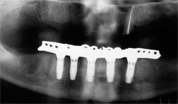 | |||||
| Figure 13 Panoramic radiograph 12 weeks post implant placement and prosthesis delivery. | |||||
| |||||


