You must be signed in to read the rest of this article.
Registration on CDEWorld is free. You may also login to CDEWorld with your DentalAegis.com account.
Historically there have been many concepts proposed for the evaluation, diagnosis, and treatment of dental occlusion. These concepts have mostly been dentally static positions, designed for bilateral balance with forces vertically aligned on the teeth.When discussing occlusion, Angle’s classification should be referenced,1 which is a concept of occlusion based on the natural dentition. Angle used the maxillary first molars as the key to occlusion. Conversely, Dawson emphasized the harmony of the maxillo-mandibular relationship, stating that there are no rigid standards of tooth alignment that in and of themselves guarantee stability or instability of the masticatory system.2
Occlusion should be considered not only as a static concept but as a dynamic one in which occlusion could affect the craniofacial and cervical systems. Normal occlusion is a product of many structural considerations and physiological processes. To have a stable dental occlusion, there must be a confluence of balanced muscle action, proper temporomandibular joint (TMJ) position, and distributed guiding contacts of teeth in the closing stroke. If the teeth do not allow the mandible to close smoothly in its trajectory, the muscles will react by erratic action, increased tension, and torquing forces that maintain the TMJ in a strained position. In the final act of closure, the point of first tooth contact takes over guidance of the mandible to full closure. If there is a discrepancy between the muscular path of closure and the initial tooth contact, the mandible will shift to fully close.
This shift of the mandible is considered to be the key to the role of occlusal interferences which cause problems. It is presently understood that all occlusal interferences do not need to be eliminated. Only those that cause a shift in positioning of the mandible need to be eliminated. Further, on full closure, teeth act as a “doorstop” for the TMJ. Therefore, missing teeth or malposed teeth may be risk factors for joint and muscle related disorders.3
In most occlusal concepts, the primary difference is one of condylar versus muscular positions. This is consistent with the textbook images of saggital condylar positions according to Posselt or the diamond shapes of the lateral excursive movements.4 However these concepts do not take into account the 3-dimensional structures of the craniofacial skeleton because of the relative difficulty of many dentists in visualizing how the muscles, TMJ, and teeth all function in harmony.
Occlusal dysfunction appears to have a direct relationship with the masticatory musculature. Imbalances in the masticatory musculature may affect the postural muscles of the head, neck, and back.5,6 Conversely, changes in head position occur in the activity in the masticato horizontal positioning of the mandible.8 Because of these occlusal-craniocervical interrelationships, the need for a clinical 3-dimensional functional assessment of the occlusion and its effect on the body is essential.
As a teaching tool for students and dentists to understand present day dental occlusion, Mehta9 first presented a simple concept of 3-dimensional assessment of maxillo-mandibular relationships. This concept is presented here as a clinical technique for any practicing dentist who wants to quickly assess a patient’s occlusion to determine possible hidden dangers in changing or correcting the existing bite.
The Occlusal Fencing Concept
The basis of the occlusal fencing concept is that “the maxilla dictates the boundaries of the mandible in final closure and is the fence within which the mandible may become trapped or remain neuromuscularly and structurally free.”9
Generally with problems related to growth and development, such as bottle feeding, mouth breathing, tonsils, adenoids, and tongue thrusting,10 the maxilla shows a deep palatal vault and a V shape indicative of an unexpanded maxilla (Figure 1). All of these problems affect the maxilla, which then affects the way the mandible fits within its confines. This is similar to the maxilla acting as a “fence” or corral in which the mandibular teeth are the sheep. If the maxilla is constricted, then the mandibular teeth will crowd to accommodate to the space allowed by the maxillary teeth. The occlusal fencing concept is based on a structural maxillo-mandibular concept rather than a dental concept of occlusion. It takes into account 3-dimensional positioning in space.
The limits of the stomatognathic system are the basis for understanding the proposed theory of occlusal fencing. As previously stated, it is the maxilla that has a problem in the growth and development because of tongue thrusting, mouth breathing, thumb sucking, and adenoids.10 The posterior part of that fence is the throat and airway space. Anything that restricts and displaces the mandible posteriorly will affect the airway space and change the head position of the patient.11
Occlusal Fence I: Anterior/Posterior Positioning
The first occlusal fence is based on the position and inclination of the maxillary incisor teeth.12 The maxillary incisors primarily dictate the anterior/posterior position of the mandible as well as the position of the mandibular condyles in their respective fossae.Depending on the position of the individual incisors and the other “fence” factors, the mandible and condyles may vary in their respective positions. The more palatally inclined the maxillary incisors, the more retruded the mandible and condyles of the TMJ.
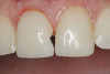
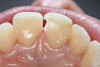
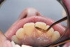
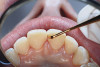
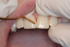
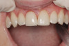
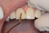
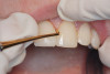
If the maxillary anterior teeth are positioned or inclined palatally, upon closure the lower teeth are going to first contact these teeth, which will force the mandible to shift to close into a retruded position. The objective is to try to identify this shift from primary contact to closure. This type of fence could potentially lead to shifting of the maxillary anterior teeth, breaking of the teeth, and/or muscular and jaw dysfunction as a result of posterior displacement of the mandible (Figure 2, Figure 3, Figure 4 and Figure 5).
The importance of the anterior/posterior positioning of the mandible has been noted previously in the literature. Dawson mentioned that disharmony between centric relation of the jaw and articulation of the teeth can cause hypersensitivity, excessive wear, and hypermobility of the teeth.2 It can also be the prime cause of pain and dysfunction of the masticatory muscles, a frequent cause of headache, as well as a cause of neck and shoulder pain.13 Jankelson reported that when centric occlusion does not coincide with the neuromuscular position, proprioceptive feedback from the mouth position in centric occlusion dictates and maintains strained muscle accommodation and an accommodated trajectory of closure.14
Occlusal Fence II: Lateral Positioning
The maxillary canines assisted by the first premolars act as the lateral positioners of the mandible and delineate the lateral fence parameters into which the mandible must finally rest. Even lateral shifts during closure as a result of interferences must ultimately succumb to the constraints of canine-to-canine space.
The position, shape, length, and inclination of the maxillary canines will affect the anterior frenal midline position of the mandible. This position may or may not coincide with the midline of the teeth, but gives an indication of the location of the left and right condyles in their fossae. The canines serve as lateral positioners of the mandible. The more lingual the maxillary canine inclination on one side, the more the mandible will shift to the opposite side and the condyle distalize on the opposite side (Figure 6, Figure 7 and Figure 8).
Fu and colleagues demonstrated that when the lateral occlusal fence is eliminated, the mandible will shift toward the midline.15 Patients with this type of fence usually present with tenderness in the lateral pterygoid. Shifting the mandible toward the midline tends to alleviate tenderness to palpation of this muscle. In addition, as the mandible moves to the midline, the condyle of the side the mandible will travel forward and medially so as to create the perception of an open bite.
Occlusal Fence III: Superior/Inferior Positioning
The concept of teeth acting as doorstops for the mandibular arch and position of the condyles requires that the multicuspid teeth be in their proper vertical height and inclination in the alveolar housing. The width of the maxillary arch during growth and development controls the positioning of the mandible. For nature to obtain the interarch tooth contact necessary for mastication, a narrow maxillary arch often leads to lingual tipping of the mandibular molars and premolars. Over time, the forces on these teeth may lead to additional tipping and consequent lowering of the vertical dimension of occlusion. This is further compounded by dental disease. Such factors allow a superior positioning of the temporomandibular condyle in the fossa as well as increased tension in the elevator muscles as a result of mechanical shortening. Patients may also develop a habit of keeping the tongue in between the teeth to maintain a relaxed vertical at rest. However during nocturnal bruxism and clenching periods, the tongue cannot rest between the teeth and it is during these periods injury may occur.
The unexpanded maxilla acts to constrain the vertical and bucco-lingual inclination of the mandibular teeth as they grow into position. This creates a reduced vertical height of that side even before aging or wear sets in and can be in the presence of all the teeth. This reduction of unilateral height tends to allow the mandible to shift toward that side especially if the first 2 fences exist. The maxillary teeth influence the height and buccal-lingual version of the mandibular posteriors. Figure 9 shows the maxilla as being the same dimension as the mandible with the mouth in a relaxed neuromuscular position. For the mandible to fit, it would have to restrict and shift posteriorly. To remain in a relaxed position, the posterior teeth must have the tongue rest between the teeth. In addition, the posterior mandibular teeth have lingually tipped in an attempt to remain in the confines of the maxilla. In cases in which this does not occur, the mandible ends up in a bilateral crossbite. Interestingly, subjects displaying class III occlusions with bilateral crossbite rarely exhibit TMJ internal derangements or loss of vertical height of the occlusion because the mandible is not constrained in any direction.
Occlusal fence III reinforces what has been suggested in the literature regarding vertical dimension. It was first suggested by Costen16 that problems within the TMJ may produce referred pain in the head and neck and that increasing the vertical dimension between the posterior teeth might reduce stress in the TMJ and thus relieve symptomatology in the referred pain areas. Gelb and Tarte emphasized that restoring occlusion to corrected vertical dimension is essential in his treatment of chronic myofascial pain dysfunction and TMJ patients.17 He recommended posterior vertical support through the use of the mandibular orthopedic repositioning appliance. This treatment claimed to be successful in alleviating headache, orofacial muscle spasms, and craniocervical pain.
Discussion
Kawamura suggested that the closing position of the jaw is primarily guided by the way the teeth fit.18 It is therefore imperative that teeth fit togther along the same closing stroke in which the TMJ and masticatory muscles function. Modifying the occlusal scheme through the use of intraoral appliances was shown by Padamsee and colleagues19 and Abdallah and colleagues20 to affect symptoms such as neck stiffness, facial pain, and headaches. It has also been suggested that mandibular position can affect the cervical spine. In a study by Aboushala and coworkers,21 vertical dimension of occlusion has been found to affect the isometric strength of the sternocleidomastoid muscle in complete denture patients. This correlation was also found in women with deep bites in whom increases in vertical dimension lead to an increase in isometric strength of the cervical flexors and deltoid muscles. Chakfa and colleagues22 demonstrated that excessive increases in vertical dimension can actually decrease isometric strength in the cervical muscles of these individuals.
The effect of posture and occlusion has been described in several studies. In an attempt to create precise occlusal schemes, much attention has been focused on the techniques of occlusal position and very little on function. As early as 1931, studies by Schwartz23 showed a relationship between gravitational posture and mandibular displacement. In 1979, Linder-Aronson and Woodside24 showed the relationship between abnormal head posture and malocclusion.
Posture has been extensively studied for upper body, shoulder, and neck problems. Kraus and Raab25 proposed a concept of musculoskeletal problems related to postural faults. Cervical problems primarily create and maintain a forward head posture,26 which has been found to affect mandibular position27 as well as airway space, tongue posture, and maxillo-mandibular growth.28,29
The 6 Keys to Normal Occlusion
When dental occlusion is evaluated and treated on a regular basis by professionals such as prosthodontists and orthodontists, it is easy to overlook the fact that dental treatment is designed to eliminate occlusal fencing and restore the occlusion to its stable and nonstrained position. For example, in 1972, Andrews presented his concept of the 6 keys to normal occlusion.30 This concept was intended as a guide to orthodontists, and it outlined 6 areas of interest in an attempt to help clinicians finish orthodontic treatment in the most ideal way. Interestingly, when all 6 keys are properly applied, correct occlusal fencing is produced.
The first key is molar relationship. In normal occlusion, the first permanent molar should be tipped slightly mesially. In a sense, this would also allow the mandible to be slightly forward, reducing the restriction in fence I. The second key is crown angulation (tipping mesially). If the maxillary teeth are in the proper angulation, the length of the upper arch would be enough to accommodate the mandibular teeth without any restrictions. The degree of incisor tip, for example, determines the amount of mesiodistal space it occupies, and therefore has a considerable effect on posterior occlusion as well as anterior esthetics. This may also affect fence I.
The third key is torque. Maxillary anterior teeth should show positive torque. This means that the root tips should be lingual while the crown tips are labial. In a sense, this position would also affect fence I, since upright upper incisors or incisors with negative torque would lead to mandibular distalization, trapping the mandible posteriorly.
When it comes to maxillary canines and premolars, this negative torque would put the maxillary teeth in a position to shift the mandible laterally, restricting fence II. Spaces in the upper arch may develop as a result of improper torque and angulation of teeth. If these spaces are closed by orthodontic treatment, it may lead to mandibular restriction (Figure 10). The fourth key is rotation. Teeth within a dental arch should assume a certain position. Mesial rotation of the maxillary first molar, for example, may lead to a false class II molar relationship, affecting the first fence and in some cases the second fence. The fifth key is tight contacts. Interproximal contacts should be tight. If there is a tooth size discrepancy, it should be corrected by using crowns, veneers, or composite buildups. Closing these spaces with orthodontics may cause the mandible to be trapped posteriorly upon maximum intercuspation. This is a fence I restriction.
The final key proposed by Andrews is the occlusal plane and the curve of spee. The occlusal plane should be flat at the completion of orthodontic treatment, allowing it to settle later on into an ideal curve. Fence III may be compromised in cases with deep curve of spee. In these situations, the vertical height of the posterior teeth is lower than normal, leading to a loss in the vertical dimension of occlusion (third fence) as well as mandibular distalization (second fence) (Figure 11 and Figure 12).
Conclusion
Clinicians evaluating and treating the dentition should keep in mind that their treatment as well as the patient’s presenting dental occlusion may have a significant impact on the TMJ, muscles of mastication, and supporting structures. Proper understanding, evaluation, and elimination of these “occlusal fences” is critical to the health of the stomatognatic system and the body as a whole.
References
1. Angle EH. Treatment of Malocclusion of the Teeth and Fractures of the Maxilla, Angle’s System. 7th ed. Philadelphia, Pa: SS White Dental MFG Co; 1990.
2. Dawson P. Evaluation, Diagnosis and Treatment of Occlusal Problems. 2nd ed. St. Louis, Mo: CV Mosby;1989.
3. Tallents RH, Macher D, Kyrkanides S, et al. Prevalence of missing posterior teeth and intraarticualr temporomandibaulr disorders. J Prosthet Dent. 2002;87(1):45-50.
4. Posselt U. Range of movement of the mandible. J Am Dent Assoc.1958;56(1):10-3.
5. Ramfjord SP, Ash MM. Occlusion. 2nded. Philadelphia, Pa: WB Saunders;1971:126.
6. Frumker SC , Kyle MA. The dentist’s contribution to rehabilitation of cervical posture and function: orthopedic and neurological considerations in the treatment of craiomandibualr disorders. Basal Facts. 1987; 9(3):105-109.
7. Funakoshi M, Amano N. Effects of the tonic neck reflex on the jaw muscles of rat. J Dent Res. 1973;52(4):668-673.
8. Lund P, Nishiyama T, Moller E. Postural activity in the muscles of mastication with subjects upright, inclined, and supine. Scand J Dent Res. 1970;78(5):417-424.
9. Mehta NR. Understanding the influence of occlusal modifiers as they affect occlusal diagnosis. KMC Dent J. 1995; 6(1):69-78.
10. Milic JD, Nikolic P, Novakovic S. Effects of adenoidectomy and immediate orthodonthic treatment on jaw relations and naso-respiratory rehabilitation. Vojnosanit Pregl. 2005;62(2):119-124.
11. Choi JK, Goldman M, Koyal S, et al. Effect of jaw and head position on airway resistance in obstructive sleep apnea. Sleep Breath. 2000;4(4):163-168.
12. Spear FM, Kokich VG, Mathews DP. Interdisciplinary management of anterior dental esthetics. J Am Dent Assoc. 2006; 137(2);160-169.
13. Fricton J, Kroening R, Haley D, et al. Myofascial pain syndrome of the head and neck: a review of clinical characteristics of 164 patients. Oral Surg Oral Med Oral Pathol. 1985;60(6):6515-6623.
14. Jankelson B. Neuromuscular aspects of occlusion : eefects of occlusal position on the physiology and dysfunction of mandibular musculature. Dent Clin North Am. 1979; 23(2):157-168.
15. Fu AS, Mehta NR, Forgione AG, et al. Maxillomandibular relationship in TMD patients before and after short-term flat plane bite plate therapy. Cranio. 2003; 21(3):172-179.
16. Costen JB. Syndrome of ear sinus symptoms dependent upon disturbed function temporomandibular joint. Ann Otolaryngology. 1934;43:1.
17. Gelb H, Tarte J. A two year clinical dental evaluation of 200 case of chronic headache: the cranio-cervical mandibular. J Am Dent Assoc. 1975;91(6):1230-1236.
18. Kawamura Y. Neurogenesis of mastication. Front Oral Physiol. 1974;1(0):77-120.
19. Padamsee M, Mehta N, White G, et al. Effect of repositioning and flat occlusal splints on masticatory muscle tenderness. Presented at: International Association of Dental Research; March 1996; San Francisco, Ca; Journal of Dental Research . 1996;75:28. Abstract 85.
20. Abdallah E, Mehta NR, Forgione A, et al. Affecting the upper extremity strength by changing maxillo-mandibualr vertical dimension in deep bite subjects. Cranio. 2004;22(4):268-275.
21. Aboushala A, Mehta N, Kugel G, et al. Vertical dimension’s effect on sternocleidomastoid strengh in complete denture patients. Journal of Dental Research. 1997; 76:142. Abstract 3185.
22. Chakfa AM, Mehta NR, Forgione AG, et al. The effect of stepwise increases in vertical dimension of occlusion on isometric strength of cervical flexors and deltoid muscles in nonsymptomatic females Cranio. 2002;20(4):264-273.
23. Schwartz AM. Prophylaxe and F Ruhandlung. Der Bissonomalian, Fortschr, Orthod. 1931;1:635.
24. Linder-Aronson S, Woodside DG. The growth in the sagital depth of the bony nasopahrynxin relation to some facial variables. In: McNamara JA, ed. Nasorespiratoy Function and Craniofacial Growth. Monograph No.9. Center for Human Growth and Development. Ann Arbor, Mi: The University of Michigan; 1979.
25. Kraus H, Raab W. Hypokinetic Disease-Diseases Produced by Lack of Exercise. Springfield, Ill: Charles C. Thomas Publisher; 1961.
26. Rocabado M, Johnston BE Jr, Blakney M. Physical therapy and dentistry: an overview. J Craniomandibular Pract. 1982;1(1) :46-49.
27. Funakoshi M, Fujita N, Takenama S. Between occlusal interferences and jaw muscle activities in response to changes in head position. J Dent Res. 1976;55:684-690.
28. Rocabado M. Biomechanical relationship of the cranial cervical, and hyoid regions. J Craniomandibular Pract. 1983;1(3):61-66.
29. Last RJ. The muscles of the head and neck: a review. Int Dent J. 1955;5:338-354.
30. Andrews LF. The six keys to normal occlusion. Am J Orthod Dentofac Orthop. 1972; 62(3):269-309.
About the Authors
Noshir Mehta, DMD, MDS, MS,FICD, FACD
Director
Craniofacial Pain Center
Professor and Chairman
Department of General Dentistry
Tufts University School of Dental Medicine
Boston, Massachussets
Emad Abdallah, BDS, MS, ABOP
Assistant Professor
Orthodontist
Tufts University School of Dental Medicine
Boston, Massachussets
Silvia Lobo-Lobo, DDS, MS
Assistant Professor
Tufts University School of Dental Medicine
Boston, Massachussets
Caroline Ceveniz, DDS, MS
Assistant Professor
Tufts University School of Dental Medicine
Boston, Massachussets
Leopoldo Correa Vasquez, BDS
Assistant Professor
Tufts University School of Dental Medicine
Boston, Massachussets