The Interdependent Relationship Between Function and Estheticsin Clinical Practice
Steven R. Acker, DDS, MS
The relationship between esthetics and function when doing prosthetic restorations has been the source of many articles and discussion in current practice. The fact that there are many clinical protocols in the treatment process has led to much controversy and confusion in both practice and the academic community.
The discussion in this article will center on a diagnostically based, decision-making protocol for achieving predictable results in prosthetic restorations. The focus will be on the first direction of treatment, followed by the modality of treatment in achieving the highest level of prosthetic restorations. The same principles will apply to all facets of prosthetic dentistry, whether it is fixed, removable, or an implant-supported prosthesis.
The first level of understanding has to deal with the intimate relationship between function and esthetics. Many outstanding clinicians have written and lectured on these concepts, including Jon Kois and others. The controversy involved in discussing occlusion and function centers on the fact that there are no standardized and accepted diagnostic entities in function and occlusion. With periodontal disease, types1 through 4 are diagnostic entities that are agreed upon throughout our profession because of the American Academy of Periodontology diagnostic protocols. When discussing occlusion and function, an agreement on diagnostic entities does not exist.1
The discussion of function needs to center around the consequences to the patient’s “chewing system” as he or she chews.1 This system is composed of teeth, muscles of mastication, and the temporomandibular joint (TMJ). When the system works in harmony, there is acceptable function. The first question is how to define acceptable function. There is a consensus that for a patient to have acceptable function, he or she cannot have age inappropriate wear of teeth, no symptoms of temporomandibular joint dysfunction (TMD), and no mobility of teeth from primary occlusal trauma.
To discuss acceptable function, some basic definitions need to be considered. These definitions appear in The Glossary of Prosthodontic Terms.2 Maximum intercuspal position (MIP), is a tooth-to-tooth position, which is determined by how teeth fit together when biting. This is independent of condylar position, and may or may not coincide with centric relation. Centric relation is the most anterior and superior position of the condyle in the articular fossa. This is strictly a jaw-to-jaw position. Finally, centric occlusion is the position of the teeth when the mandible is in centric relation.3
The next step in the clinical decision-making process is establishing a functional record for the restorative or prosthetic treatment. If the patient is truly in acceptable function, tooth to tooth position can be used in fabricating restorations. An MIP record, using a polyvinyl siloxane (PVS) bite registration material will provide an acceptable functional record for this patient.
Although some patients will fit into the acceptable function group, many patients present with areas of their “chewing system” that are not in acceptable function. The question then becomes, “What clinical decision-making protocol should be used to restore these patients?” Many of these patients have significant destruction of tooth structure from improper loading. A single tooth or multiple teeth may prevent the proper seating of the condyle. The teeth are in the “wrong place” for all practical purposes. The result of this type of dysfunction may be age inappropriate wear to tooth structure or TMD symptoms. The chewing system is not working in harmony. If the restorative system is rebuilt to its previous position, the prosthesis or restoration will be at risk for the same wear and destruction that occurred to the natural tooth. This time it will be a prosthetic failure, but the mode of failure may be different. Failure of the prosthesis/restoration may be from cement fatigue or porcelain fracture.
When it has been determined that the teeth are in the “wrong place,” a predictable method of restoring the tooth or teeth to the “right place” needs to be created. At this point, centric relation needs to be used as our fall-back position to restore the patient.4 The first step in the decision-making protocol is to determine if centric relation is needed. This is the fall-back position, not the driving force in our clinical decision making.
The chewing system in these patients has shown destruction for reasons dealing with teeth, TMD, and/or musculature.5 However, between 5% and 8% of patients have neurologic factors as the etiology of destruction of teeth.6 These patients are those with true nocturnal bruxism, and show linear wear patterns on all of the occluding teeth. This can be confirmed by examining occlusal nightguards of these patients for linear streaks and seeing previous wear patterns of the teeth. Patients with certain neurologic diseases and some drug-induced responses are included in this group.7 The clinical decision-making protocol for these patients deals with minimizing anterior guidance and restoring them to control the destruction caused by the central factors.
The majority of patients with functional disease are the result of disparities of the chewing system as described. Kois has described these patients whose anterior teeth have frictional contact when chewing as the first sign of wear as having a constricted chewing pattern.6 In effect, these patients contact their incisors before the posterior teeth when chewing. It is the anterior teeth that prevent or are the first point of contact before the condyle seats. The “envelope of function,” as described by Dawson, is too tight.3,4 Typical wear patterns in these patients are the lingual incisal of the maxillary anterior teeth, or the labial incisal of the mandibular anterior teeth (Figure 1). Conversely, when the initial points of contact when chewing are the cusps or cuspal inclines of posterior teeth, destructive wear patterns are often seen on these teeth. In past literature, these were often called prematurities.3 Kois has described these patients as having occlusal dysfunction as their functional diagnosis.6
By categorizing patients who have no central or neurologic etiology for functional risk as acceptable function, constricted chewing pattern, or occlusal dysfunction (as defined by Kois), a terminology has been created that all practitioners in all disciplines of dentistry can refer to. A central terminology helps restorative dentists in multidisciplinary cases communicate with orthodontists, periodontists, oral surgeons, or anyone placing implants. It is a basis for the standardization of functional diagnosis in dentistry.
Once a functional diagnosis has been established, the next level of decision making in restoring the patient to maximum esthetics and function needs to be accomplished. When a patient is in acceptable function, there is no need to find a centric-relation position. An MIP record using any PVS bite registration material can be used.
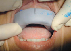
A face-bow record can be taken to translate the position of the maxilla (Figure 2). The maxillary incisal edge position can now be determined. This can be accomplished using esthetic criteria to determine the vertical position of the maxillary incisal edges as the first level of determining esthetic position. Digital photographs of the patient in repose and in “smile” position can be used. The smile position can be determined by asking the patient to say “EE.” The laboratory should be instructed to create a wax-up for the case by using a vertical position of the patient showing 2mm of the central incisor in repose (Figure 3). When designing a full case, the laboratory should be instructed to create the maxillary posterior occlusal plane, followed by the mandibular incisal edge position, and finally, the mandibular posterior occlusal plane. In acceptable function, a single tooth restoration can be designed if indicated.
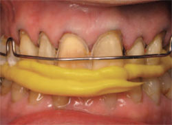
If the functional diagnosis is a constricted chewing pattern or occlusal dysfunction, the centric-relation position is needed to properly restore the patient, and have the teeth, TMJ, and musculature working in harmony. A dependable, reproducible, and accurate methodology for determining centric relation is the use of a Kois Deprogrammer (Creating Restorative Excellence, Inc, Seattle, WA) (Figures 4 and 5). The discluding element just posterior to the maxillary central incisors allows the condyle to seat in the superior and anterior aspect of the fossa. It does this by taking the teeth out of the equation. The only point of contact is the mandibular incisors on the discluding element of the deprogrammer. Adjusting the deprogrammer to 1 point on the discluding element after 1 week, and taking a centric-relation record after 2 additional weeks will allow a restoration design that will bring the patient to acceptable function.8
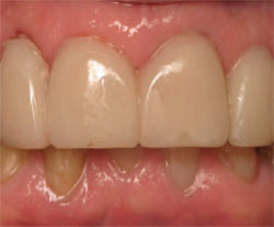
The centric-relation record taken with the deprogrammer in place as well as a face-bow record is sent to the laboratory (Figures 6).9 In our practice, the face-bow/articulator system used is the Panadent® System (Panadent Corporation, Grand Terrace, CA). The laboratory can then create a wax-up for the prescribed restorations. The goal of function is to have simultaneous, bilateral, equal intensity contact on cuspids and the supporting cusps of bicuspids and molars. The incisors should not create any friction in this position. A PVS matrix can be fabricated on the final wax-up (Figure 7). This will allow provisional restorations to be fabricated at the final impression visit (Figures 8 and 9). These restorations are used to create ideal function and esthetics for the patient.6 They are truly trial restorations. The vertical position of the restorations is determined by esthetics, and the horizontal position of the restorations is driven by the functional decisions.
CONCLUSION
The described diagnostic and treatment protocols provide a predictable protocol for the intimate relationship between esthetics and function in all aspects of restorative and prosthetic dentistry. Centric relation is used as a fall-back position in patients who are not in acceptable function. This position is reliably determined by the use of a Kois Deprogrammer. The information is transferred to the laboratory by the use of our Panadent face bow and the centric-relation record of the deprogrammed patient.
REFERENCES
1. Dawson PE. Optimum TMJ condyle position in clinical practice. Int J Periodontics Restorative Dent. 1985;5(3):10-31.
2. The glossary of prosthodontic terms. J Prosthet Dent. 2005;94(1):10-92.
3. Dawson PE. A classification system for occlusion that relates maximum intercuspation to the position and condition of the tempromandibular Joints. J Prosth Dent. 1996;75(1):60-66.
4. Dawson PE. New definition for relating occlusion to varying conditions of the temporomandibular joint. J Prosthetic Dent. 1995;74(6): 619-627.
5. McNeill C. The optimum tempromandibular joint condyle position in clinical practice. Int J Periodontics Restorative Dent. 1985; 5(6): 52-76.
6. Kois J. 10 Step Management Approach, Creating Restorative Excellence, Center for Advanced Learning Seminar Series.
7. Gelb H. The optimum tempromandibular joint condyle position in clinical practice. Int J Periodontics Restorative Dent. 1985; 5(4): 34-61.
8. Long JH Jr. Diagnostic tests used in determining the role of occlusion in tempromandibular joint disorders. J Prosthetic Dent. 1991;66(4):541-544.
9. Long JH Jr. Occlusal adjustment as treatment for tenderness in the muscles of mastication in category 1 patients. J Prosthetic Dent. 1992;67(4):519-524.
10. Long JH Jr, Buhner WA. New diagnostic and therapeutic mechanical devices. J Prosthetic Dent. 1992;68(5): 824-828.
11. Long JH Jr. A device to prevent jaw clenching. J Prosthetic Dent. 1998;79(3):353-354.
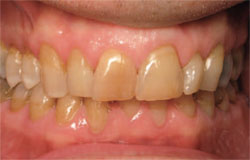 | 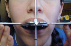 | ||||
| Figure 1 When the anterior teeth prevent (or are) the first point of contact before the condyle seats, typical wear patterns are the lingual incisal of the maxillary anterior teeth or the labial incisal of the mandibular anterior teeth. | Figure 2 A face-bow record can be taken to translate position of the maxilla. | ||||
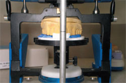 | 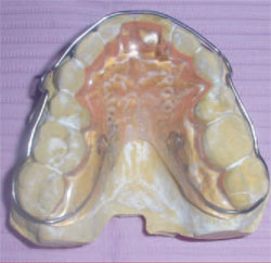 | ||||
| Figure 3 A wax-up can be created by using a vertical position of the patient, which shows 2 mm of the central incisor in repose. | Figure 4 The Kois Deprogrammer. | ||||
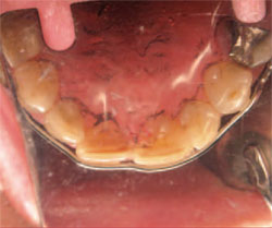 |  | ||||
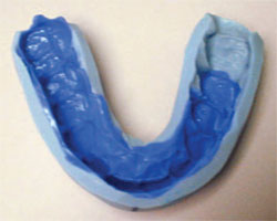
| Figure 5 The Kois Deprogrammer placed in the maxilla. | Figure 6 A centric-relation record is taken with the Deprogrammer in place. | ||||
 |  | ||||
| Figure 7 A PVS matrix can be fabricated on the final wax-up. | Figure 8 Taking the final impression. | ||||
 | |||||
| Figure 9 The final restorations. | |||||
| |||||