
An Articulated Discussion About Articulators
Lee Culp, CDT
Articulators are a big topic of discussion these days among clinicians and laboratory technicians because their usefulness and necessity in the treatment of functional, esthetic, and occlusal problems has become more significant. Although I can’t provide specific recommendations for which articulator system is best for your situation, I can describe the different articulator categories available and outline some options to consider before purchasing a system.
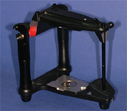
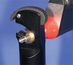
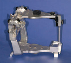
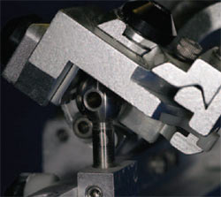
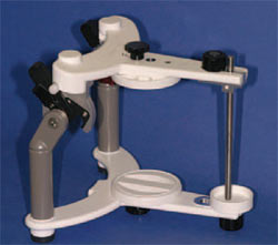
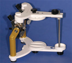
Basically, articulator systems are divided into 2 groups: Arcon and non-Arcon. Arcon articulators feature a mechanical condyle located on the lower frame of the articulator that imitates the condyle of the natural joint (Figures 1 and 2). The joint cavity imitating the natural joint fossa is part of the upper frame of the articulator. Non-Arcon articulators place the mechanical condyle on the upper part of the frame.1 In other words, the Arcon articulator copies the natural bones of the skull, while the non-Arcon articulator is in reverse, with the condyle moving in the opposite direction (Figures 3 and 4).
Non-anatomical Articulators
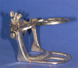
There are also non-anatomical articulators, which do not accurately represent anatomical movements of the mandible as a result of their small size.2 Referred to as an occluder or, in dental slang, a “barn-door hinge”, this type of articulator only permits opening and closing movements around a fixed axis (Figure 5). Lateral, protrusion, or retrusion movements cannot be accurately achieved. Model orientation using non-anatomical articulators is arbitrary and does not provide a representation of the natural relationship to the patient’s temporomandibular joint (TMJ). Because no relationship to the TMJ can be established and no excursive movements can be simulated, these articulators are best suited for small single-unit cases in which excursive movements can be duplicated from the wear patterns on the remaining teeth.
Anatomical Articulators
However, as a result of extensive research and development, anatomical articulators are now available that can very accurately replicate anatomical movements of the mandible. Within this category are 3 primary types.3-5
Average-value Articulators are equipped with an average protrusive path of approximately 30°. Therefore, setting individual patient parameters is not possible because the condyles are fixed. Model orientation is generally accomplished according to average values5 (Figure 6).
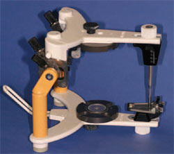
Semi-adjustable Articulators usually permit the transfer of individual patient parameters, such as the inclination of the protrusion path and the Bennet angle. Using a facebow transfer for model orientation, a relationship to an individual patient’s TMJ is possible (Figure 7).
Fully-adjustable Articulators enable all of the above mentioned settings, in addition to individual adjustable settings.4 These include inter-condylar, programmable retrusion, and custom settings to duplicate the exact condylar movements of a specific patient. Settings on fully-adjustable articulators are programmed using a manual or computer-generated pantographic tracing6,7 (Figure 8).
Considerations for Selection
Aside from the mechanics, there are several other variables to consider before deciding which articulator system to purchase.
Price. The more complex the articulator becomes, obviously the higher the price. Generally, average-value articulators range in price anywhere from $100 to $600, depending upon the quality of the instrument and the accessories offered for the system. Semi- and fully-adjustable articulators can range in price from $500 to more than $1,500, with pantographic instrumentation costing up to $5,000.
Ease of Use. Choose an articulator that is “technician friendly”. Many on the market today are excellent diagnostic tools but do not hold up very well under the rigors of everyday use. A feature I find necessary in an articulation system is increased vertical height between the upper and lower mounting structures. Low vertical-height systems sometimes will not allow correct facebow positioning of the model when both the upper and lower arches are pinned, because each requires too much space.
Calibration. When dentist/technician teams choose an articulator system, calibration between their respective articulators becomes a necessity. Calibration allows models from one articulator to be accurately transferred to another. When working with laboratories outside of your geographical area, calibration transfer enables models to be shipped without sending your articulator. Transfer becomes even easier if the articulator offers the option of a magnetic mounting system, which is becoming very popular because it facilitates quick and efficient model transfer between the dentist and the laboratory.
Conclusion
Choosing an articulator system should be a combined effort between the clinician and the laboratory. Take your time when researching all of the systems available and, ultimately, choose the one(s) that will best suit your needs. The initial cost may seem expensive, but purchasing a quality articulator system is a worthwhile investment for producing the high-quality dentistry that’s becoming more and more in demand.
References
1. Kubein-Meesenburg PD, Meyer DG, Bucking DW. Practical application of an anterior guidance reconstruction concept. 2. Clinical use of the C.C.F. Cah Prosthese. 1990;69:36-42.
2. Hobo S, Shillingburg HT Jr, Whitsett LD. Articulator selection for restorative dentistry. J Prosthet Dent. 1976;36(1):35-43.
3. Rihani A. Classification of articulators. J Prosthet Dent. 1980;43(3):344-7.
4. de Carvalho Ode T. A new fully adjustable articulator system and procedure. J Prosthet Dent. 1998;80(3):376-86.
5. Ecker GA, Pameijer CH. Comparison of the Panadent Quick Analyzer and Analog Selector systems. Int J Prosthodont. 1989;2(6):531-6.
6. Mandilaris CB, Beard CC, Clayton JA. Comparison of the intercondylar distance and the interfacial width as used with the electronic pantograph. J Prosthet Dent. 1992;67(3):331-4.
7. Hatano Y, Kolling JN, Stern N, et al. A graphic comparison of mandibular border movements generated by various articulators. Part II. Results. J Prosthet Dent. 1989;61(4):425-9.
 |
 |
|
| Figure 1 An Arcon articulator. | Figure 2 Close-up of the condyle assembly on an Arcon articulator. | |
 |
 |
|
| Figure 3 A non-Arcon articulator. | Figure 4 Close-up of the condyle assembly on a non-Arcon articulator. | |
 |
 |
|
| Figure 5 A non-anatomical articulator. | Figure 6 Example of an average-value articulator. | |
 |
 |
|
| Figure 7 Example of a semi-adjustable articulator. | Figure 8 Example of a fully-adjustable articulator. | |
| About the Author | ||
 Lee Culp, CDT Lee Culp, CDTMosaic Studios, Inc. Institute for Oral Art and Design Bradenton, FL www.ioadsmiles.com |
||

