Changing Tooth Position with Orthodontics or Restorative Dentistry: Both Perspectives
Paul R. Chalifoux, DDS; Steve Noxon, DMD, MSD
Dentistry often provides multiple treatment options for patients. The treatment planning process is difficult because various options present advantages and disadvantages for individual patients and, therefore, must be customized for each case. Tooth replacement is a common example, as it can be accomplished with removable partial dentures, fixed bridges, or implants. The appropriate treatment is based on constraints such as financial limitations, patient preferences, long-term prognosis, patient comfort, physical barriers, psychological considerations, and the dentist’s and laboratory’s ability to perform the work. Treatment planning to correct misaligned teeth is similar.
Correcting the appearance of misaligned teeth is accomplished with orthodontics, restorative dentistry, or both. Tooth misalignment includes tooth rotation, crowding, super-eruption, under-eruption, intrusion, diastemas, and labial, lingual, mesial, or distal tipping. Misalignment also occurs from inappropriate arch shape, position and size, and arch-to-arch relationships. Correction with orthodontics or restorative dentistry presents limitations, advantages, and disadvantages.
Restorative Techniques and Limitations
It is important to understand what can be achieved with restorative dentistry, orthodontics, and combined treatments. Restorative dentistry changes tooth morphology by removing tooth structure or adding restorative materials to replace missing tooth structure and increase dimensions. Changes are made to the coronal portion of a tooth without movement of the root. The root dimensions at the cemento-enamel junction or where a root exits the gums and alveolus cannot be moved restoratively and, therefore, become a limiting factor. Orthodontics is required to change root position. Consequently, maximum and minimum tooth width is determined by the root position of a tooth and the adjacent teeth.
Tooth reduction is a second limiting factor. Removal of tooth structure must leave sufficient support to resist forces without fracturing. For example, a lateral incisor that has a small cross section following crown preparation can fracture through dimension, leaving the coronal portion of a tooth in a crown. Reduction cannot encroach on the pulp or it will lead to pulpal necrosis unless endodontic treatment is planned.1-5 Removal of enamel that exposes dentin must allow enough room to restore over dentin to prevent sensitivity.2,6-8
Restorative Illusions
Changes in tooth anatomy without altering overall tooth dimensions can create an appearance of tooth movement. Controlling the face and silhouette of a tooth provides visual indicators of perceived size and long axis orientation. A large tooth looks smaller when the face is small, while the silhouette fills the space, thereby not requiring a change in existing tooth-to-tooth space.9-12
The face of a tooth is defined as the central portion of a facial surface within the mesial and distal line angles, the gingival height of contour and the incisal edge or transitional incisal curvature.9-12 The silhouette is defined by the outside edges of the tooth, including the gingival contour, gingival and incisal embrasures, contacts, and incisal edge.9-12
The face of a tooth looks smaller as line angles, gingival height of contour, and incisal curvature approach the center of a tooth.9-12 Curved line angles, combined with increased embrasure size, reduce the incisal length to create a smaller appearance. Color variation, as well as surface contour and texture of the face of a tooth, make teeth look smaller, wider, or longer.9-12 For example, 2 vertical grooves between lobes of a central incisor create a longer looking tooth, while horizontal grooves make a central incisor look wider. Ideally, placement of grooves or surface texture should replicate naturally occurring patterns.
Case #1
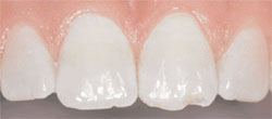
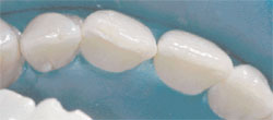
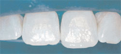
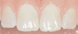
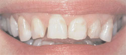
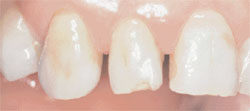
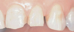
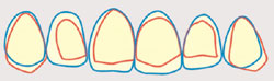
An incisal fracture and chipping on the left central incisor require restoration to recreate proper anatomy (Figure 1). Further evaluation reveals rotation of the tooth (Figure 2 ). Composite is added to the facial surface and restores the incisal edge. Increased thickness at the incisal edge from increasing the facial dimension creates a more durable restoration and reduces the appearance of malalignment/ rotation (Figure 3 ). This tooth could have been rotated orthodontically, but doing so would not have rendered a superior esthetic result (Figure 4) and would have compromised the durability of the final restoration by under-utilizing the bonding surface on the facial aspect.
Restorative Treatment Planning
Improving smiles takes into consideration 3 major defect categories: tooth position, color, and loss of tooth structure. Changing the appearance of tooth position with restorative dentistry may be preferable to orthodontics if restoration is required to correct color flaws or loss of tooth structure.13 Tooth color defects include discoloration, stains, fluorosis, tetracycline stains, decalcifications, hypocalcifications, craze lines, fillings, root exposure, and cavities. Tooth structure losses include chips, cracks, abrasion, attrition, surface defects, exposed root structure, surface contour defects, surface texture defects, factures, cavities, and developmental defects such as amelogenisis or dentiogenisis imperfecta.
When treatment planning, existing tooth position is compared to ideal tooth position. Tooth structure outside the ideal position requires tooth reduction. If the magnitude of reduction extends beyond enamel into sensitive dentin, extra reduction is required so that a restoration can cover the dentin surface. Teeth require restoration to add tooth mass if the ideal tooth position is outside of the existing position.14, 15 Drawing teeth 2-dimensionally from an occlusal or facial perspective can help develop the restorative approach. The technique of drawing is effective for very experienced dentists who can envision objects 3-dimensionally from a series of 2-dimensional images.
Diagnostic wax-ups create 3-dimensional images and predictable results. Wax is added to areas that require increased dimension, and the diagnostic models are marked to indicate areas of reduction. Alternatively, a duplicate model can be trimmed extensively, and the teeth can be waxed to full contour. A visual comparison is made of the original study model and the ideal waxed model. An impression of a waxed model is poured in stone, and a clear vacuum form is used as a template to verify treatment progress intra-orally.
Case #2
In this unaesthetic smile (Figure 5), the teeth have dark and irregular color patterns. Wear has reduced incisor length, chipped teeth unevenly and developed irregular facial contours. Loss of posterior support has caused the maxillary cuspids to flair buccally (Figures 6 and 7). There are minor irregularities in the gingival contours, especially around the right central incisor.
A drawing of the existing tooth shape and position in red, with an ideal tooth position and shape in blue, was rendered (Figure 8). Areas where blue lines are outside of red lines required the addition of tooth structure or gingival recontouring, while areas defined by red lines outside of blue lines required reduction. Three dimensionally, the cuspids required extra preparation to reduce the buccal flair, while the lingual tipped incisors required less reduction and additional material to produce a more buccal position.
In this case, the patient’s teeth required restoration for defects in tooth color and structure (Figure 9), and it was determined that correction of the appearance of tooth position could be accomplished at the same time. Evaluation of the primary 2 limiting factors of restorative treatment alone (i.e., root position and the amount of remaining tooth structure) revealed that orthodontics was not required to supplement treatment. Full-mouth reconstruction without braces resulted in a perfect smile (Figure 10).
The Orthodontic Perspective
The ability to physically move teeth presents myriad possibilities that could substantially improve many patients’ appearances. When an orthodontist evaluates a case for treatment, he/she is considering a variety of functional and esthetic goals. First and foremost, an orthodontist looks at the basic functional guidelines learned in dental school: overjet (i.e., how much the top teeth flare labially from the bottom teeth); overbite (i.e., how much vertical overlap exists between the front teeth); transverse relationship between upper and lower teeth; facial profile; crowding/spacing; excursive movements; etc. It is oftentimes an aberration in these basic guidelines that prompts a referral to an orthodontist. These are well understood principals; but what about the more subtle concepts? What can an orthodontist do for patients who have some restorative needs or are simply displeased with the esthetic appearance of their smiles?
The first thing all orthodontists should do in an examination is look at a patient’s smile. It sounds obvious, but it is rarely done. Patients should smile naturally and laugh so they can be viewed very critically. Does the patient show any gingiva? Are the patient’s gingival margins esthetic? Is the patient’s maxillary dental midline coincident with their facial midline? Do the patient’s maxillary incisal edges follow the contour of their lower lip? Are there black spaces in between the contact points of adjacent teeth and the papilla? Do the teeth look symmetric and proportional vertically and horizontally? Are the patient’s buccal corridors filled with nicely colored tooth structure or black cavernous spaces? Are there large spaces, areas of crowding, tipped teeth, or unevenly worn teeth? These are all common findings that an orthodontist has the ability to manipulate.
If a patient shows gingival tissue when smiling, it is equally important to treatment plan gingival esthetic goals because it is a dental esthetic goal. Zero to 2.0 millimeters of gingival display is highly esthetic and appears youthful.16, 17 As a general rule, a patient’s gingiva should appear 0.5 to 1.0 mm higher on the central incisors and canines than the lateral incisors.18 Most importantly, however, similar classes of teeth should appear symmetric bilaterally.19 Gingival margins that do not follow this general guideline appear uneven, thereby making some teeth appear too high/low or disproportionately long/short. In the absence of periodontal disease or any other complicating factors, the gingival margins will follow teeth as they are moved and afford an orthodontist the opportunity to manipulate their positions as they reposition teeth.20
Of the many complicating factors impacting an orthodontist’s ability to achieve perfectly esthetic gingival margins are variations in tooth proportions. This is a very common problem among adult patients because malaligned teeth are usually worn unevenly. When this is the case, an orthodontist, working in conjunction with a restorative dentist, should evaluate tooth proportions to determine whether the longer adjacent teeth should be recontoured at their incisal edge to match the worn tooth, or the worn tooth built up to match the longer, unworn contralateral tooth. That determination is commonly based on the rule of golden proportions that outlines the appropriate sizes of adjacent teeth in an arch.21-24
Using a central incisor as a frame of reference, we know that the ideal width-to-height ratio in most faces is between 75% to 80%.25, 26 If, for example, a central incisor is 8.75 mm wide, its height should be between 11.7 mm for a longer appearing tooth and 10.9 mm for a shorter appearing tooth. If the gingival margin of this 8.75-mm wide tooth is at 1.5 mm incisal to the ideal gingival margin position, and the unworn adjacent tooth is sized according to the dimensions above, we would intrude this tooth to achieve esthetic gingival margins and build up its height to the properly sized adjacent central incisor. If, on the other hand, the adjacent unworn central incisor was more than 12.4 mm (e.g., 10.9 mm plus 1.5 mm), we would extrude the unworn central incisor to achieve esthetic gingival margins and enamelplasty its incisal edge so that it matches the proportions of the worn, but proportionately sized, adjacent central incisor. Naturally, some situations warrant a combination of both approaches.
Once the ideal gingival margin locations and tooth proportions have been identified, teeth can be positioned to distribute spaces ideally for restorations. Ideally positioned tooth preparations will allow for more uniform tooth reduction and restoration; decreased areas of heavy reduction and subsequent pulp exposures with future maintenance restorations; decreased periodontal complications from overbulked restorations; and highly esthetic emergence profiles.27 If, for example, space is needed for restoration of a pegged lateral incisor, it should be positioned so that two-thirds of the space is distributed on the distal side of the tooth and one-third of the space is distributed on the mesial side.27 If this same tooth is positioned with the majority of the space on the mesial, this side will be overbulked and generate an overly acute emergence profile. Remember the rule about gingival margin levels? It is also imperative that pegged laterals be positioned vertically to generate a gingival margin that is symmetric with the contralateral incisor gingival margin.
Unfortunately, even the best sized and proportioned teeth can still look awkward if the midline of the maxillary teeth does not align with the middle of a patient’s face.
Treatment of a non-coincident midline greater than 1 mm would involve physically moving teeth orthodontically. Very large movements may even involve the reduction of tooth mass using interproximal reduction or asymmetric extractions.
Additionally, well-aligned teeth do not look natural if their incisal edges do not follow the contour of a patient’s lower lip. Maxillary arches that appear linear transversely look very artificial. The vertical dimensions of the maxillary incisors, however, cannot be augmented too much before they stand in violation of generally accepted esthetic tooth proportions and appear very long and slender.21-26 That is, of course, unless that patient has inadequate incisal display when they smile and hides the excess crown length behind their upper lip. It is imperative, therefore, that orthodontists position teeth in a way that is vertically harmonious with the shape of a patient’s lower lip when they smile.
A maxillary arch that follows the mandibular lip contour should also diminish into a patient’s cheeks and not appear to end abruptly distal to the patient’s canines.28 This objective is loosely referred to as a broad smile or narrow smile, but more precisely described by a qualification of buccal corridor volume. Treatment of a patient with large buccal corridors has to proceed cautiously, because expanding the transverse dimension of the maxillary arch so that it is disproportional with the mandibular arch would introduce a significant functional compromise. Additionally, this movement in a patient with a resistance at the mid-palatine suture would merely tip teeth buccally and, consequently, be unstable and potentially detrimental to long-term gingival health (recession).29
Conclusion
Changing tooth position may involve and take into consideration orthodontics and/or restorative options. Thorough diagnosis of tooth position, structure, and color are required for treatment planning. Patient goals and limitations to treatment define expected results, as well as the most appropriate option selected.
References
1. King M. Preserving the vital pulp in operative dentistry. Dent Update. 2003;30(3):159.
2. Tecles O, Laurent P, Zygouritsas S, et al. Activation of human dental pulp progenitor/stem cells in response to odontoblast injury. Arch Oral Biol. 2005;50(2):103-8.
3. Vij R, Coll JA, Shelton P, et al. Caries control and other variables associated with success of primary molar vital pulp therapy. Pediatr Dent. 2004;26(3):214-20.
4. Bichacho N, Eylat Y, Dadi M, et al. Restoration modalities of severely injured anterior teeth--gingival integration, papillae support, and predictable imperfections. Compend Contin Educ Dent. 2003;24(12):891-4, 896-8, 900-4.
5. Scarano A, Manzon L, DiGiorgio R, et al. Direct capping with four different materials in humans: histological analysis of odontoblast activity. J Endod. 2003;29(11):729-34.
6. Magne P. Immediate dentin sealing: a fundamental procedure for indirect bonded restorations. J Esthet Restor Dent. 2005;17(3):144-54.
7. Bassi GS, Youngson CC. An in vitro study of dentin exposure during resin-bonded fixed partial denture preparation. Quintessence Int. 2004;35(7):541-8.
8. Lockard MW. A retrospective study of pulpal response in vital adult teeth prepared for complete coverage restorations at ultrahigh speed using only air coolant. J Prosthet Dent. 2002;88(5):473-8
9. Chalifoux PR. Checklist to aesthetic dentistry. Pract Periodontics Aesthet Dent. 1990;2(1):9-12.
10. Singer BA. Principles of esthetics. Curr Opin Cosmet Dent. 1994;6-12.
11. Singer BA. Fundamentals of esthetics. In: Dale BG, Aschheim KW, eds; Esthetic Dentistry: A Clinical Approach to Techniques and Materials. 1st ed. Philadelphia: Lea & Febiger; 1993:5-13.
12. Chalifoux PR. Perception aesthetics and light-cured composites. Pract Periodontics Aesthet Dent. 1992;4(5):51-56.
13. Chalifoux PR. Perception esthetics: factors that affect smile design. J Esthet Dent. 1996;8(4):189-92.
14. Magne P, Belser UC. Novel porcelain laminate preparation approach driven by a diagnostic mock-up. J Esthet Restor Dent. 2004;16(1):7-16.
15. Morgan DW, Comella MC, Staffanou RS. A diagnostic wax-up technique. J Prosthet Dent. 1975;33(2):169-77.
16. Chiche GJ, Pinault A. Esthetics of Anterior Fixed Prosthodontics. Chicago: Quintessence Pub. Co.;1994.
17. Kokich VO Jr, Kiyak HA, Shapiro PA. Comparing the perception of dentists and lay people to altered dental esthetics. J Esthet Dent. 1999;11(6):311-24.
18. Rufenacht C. Structural esthetic rules. In: Rufenacht CR, ed. Fundamentals of Esthetics. Chicago: Quintessence Pub. Co.; 1990:67-135.
19. Kokich VG, Spear FM. Guidelines for managing the orthodontic-restorative patient. Semin Orthod. 1997;3(1):3-20.
20. Kokich VG, Kokich VO. Interrelationship of orthodontics with periodontics and restorative dentistry. In: Nanda R, ed. Biomechanics and Esthetic Strategies in Clinical Orthodontics. St. Louis: Elsevier Saunders; 2005:348-373.
21. Lombardi RE. The principles of visual perception and their clinical application to denture esthetics. J Prosthet Dent. 1973;29(4):358-82.
22. Levin E. Dental esthetics and the golden proportions. J Prosthet Dent. 1978; 40:244-252.
23. Brisman AS. Esthetics: a comparison of dentists’ and patients’ concepts. J Am Dent Assoc. 1980;100(3):345-352.
24. Qualtrough AJ, Burke FJ. A look at dental esthetics. Quintessence Int. 1994;25(1):7-14.
25. Gürel G. The Science and Art of Porcelain Laminate Veneers. London, Chicago: Quintessence; 2003.
26. Gillen RJ, Schwartz RS, Hilton TJ, et al. An analysis of selected normative tooth proportions. Int J Prosthodont. 1994;7(5):410-7.
27. Kokich VG. Managing orthodontic-restorative treatment for the adolescent patient. In: McNamara JA Jr, Brudon WL, Kokich VG, eds. Orthodontics and Dentofacial Orthopedics. Ann Arbor: Needham Press, Inc.; 2001:395-422.
28. Sarver DM. Principles of cosmetic dentistry in orthodontics: Part 1. Shape and proportionality of anterior teeth. Am J Orthod Dentofacial Orthop. 2004;126(6):749-53.
29. Carmen M, Marcella P, Guiseppe C, et al. Periodontal evaluation in patients undergoing maxillary expansion. J Craniofac Surg. 2000;11(5):491-4.
Note:
All photographs are published with permission from www.dentalcomposites.com.
 |
 |
|||||
| Figure 1 Preoperative view of a patient with an incisal fracture. | Figure 2 Further evaluation reveals tooth rotation. | |||||
 |
 |
|||||
| Figure 3 View of the composite build-up prior to surface contouring and texturing, incisal edge characterization, and polishing. | Figure 4 The final restoration demonstrating how tooth anatomy, color, and position blend with the surrounding teeth. | |||||
 |
 |
|||||
| Figure 5 Preoperative view of a patient with excessive wear, chipped teeth, and irregular facial contours. | Figure 6 View demonstrating how loss of posterior support has caused the maxillary right cuspid to flare buccally. | |||||
 |
 |
|||||
| Figure 7 Alternate view demonstrating the same effect on the maxillary left cuspid. | Figure 8 A 2-dimensional drawing of the existing tooth shape and position in red, with the ideal position and shape in blue, to determine areas requiring reduction and addition of mass. | |||||
 |
 |
|||||
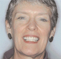
| Figure 9 Preoperative full-facial view. | Figure 10 Postoperative full-facial view. Evaluation of 2 primary limiting factors of restorative treatment alone revealed that orthodontics was not required to achieve this exceptionally esthetic result. | |||||
| About the Authors | ||||||
|
||||||