You must be signed in to read the rest of this article.
Registration on CDEWorld is free. You may also login to CDEWorld with your DentalAegis.com account.
Properly conducted orthodontic treatment enables oral and maxillofacial surgeons to obtain the best possible skeletal correction with optimal dentofacial esthetics and long-term occlusal function. Astute orthodontic preparation will likely minimize total anesthesia time by allowing for a shorter and highly precise surgical procedure. Biomechanics in surgical preparation are usually contradictory to conventional orthodontics, astooth movement is performed in the opposite direction.1
For any comprehensive surgical-orthodontic treatment, it is important that clinicians address patient concerns and understand the patient's values and expectations, taking into account the patient's motivation and psychological reasons for the treatment.2 Orthodontic and surgical computer software for simulation can play a key role in educating patients on their dentofacial deformity and possible need for combined solutions.
Adult patients with dentofacial deformities do not have the anatomical and physical growth that children and adolescents do. Therefore, an orthodontist may need the contribution of an oral and maxillofacial surgeon to correct severe skeletal problems in different planes of space that affect function, facial esthetics, and even self-awareness in adult patients.1,3 Another difference versus children and adolescents is that adults often require prosthodontic and periodontal interdisciplinary treatment.
Diagnosis: Elements to Evaluate
Surgical-orthodontic cases are diagnosed and treated in three planes of space to accomplish treatment goals.1,3-5 The following elements are evaluated by the orthodontist along with the oral and maxillofacial surgeon:
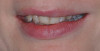
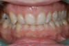
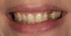
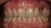
Facial thirds. First, the facial thirds need to be established. A thorough examination of the lower facial third itself is required to determine if there is a need for a lower facial height alteration. This evaluation, involving mostly the orthodontist, also includes the width of the nose, position of the ears, projection of the forehead, and the midface. Frontal, lateral, and profile photographs and images complement the clinical examination (Figure 1 through Figure 5).1,3
Symmetry of the face. In asymmetries, one of the first considerations should be the buccolingual torque of posterior teeth.6 Factors also include whether the upper, lower, and facial midlines are well-aligned1,3,6 and whether asymmetric growth has occurred. The clinician should ascertain if the patient received trauma to the chin with a condylar fracture as a youth. In many asymmetries the height of the orbits will be altered, which can also raise concerns.
Transverse dimension. The transverse dimension is the foundation for vertical and sagittal corrections.4,5 The diagnosis of maxillary transverse deficiencies is made by analyzing the posteroanterior cephalometric x-ray according to the Ricketts analysis7 or the Penn transverse analysis on a cone-beam computed tomography (CBCT) scan.8 Norm of maxilla minus mandible should be a positive measurement of 5 mm8 instead of only diagnosing the patient based on the articulated dental casts or intraoral examination.3 Skeletal assessment is used to determine the amount of surgical expansion required to resolve the discrepancy between the widths of the mandible and maxilla if present.8
Vertical dimension. Vertical dimension assessment from profile and frontal photographs and the lateral cephalometric x-ray (Figure 6) is also critical. Clockwise or counterclockwise mandibular rotations influence the sagittal position of the lower jaw. When considering vertical maxillary excess, the amount of upper incisor display with lips at rest and while smiling can be a concern. Factors may include the presence of a "gummy smile," supra-eruption of the maxillary anterior teeth, or an altered passive eruption.9 It must be determined whether bones (maxilla), the upper lip, gingival tissue, or overerupted maxillary anterior teeth are responsible for vertical problems.
Sagittal position. The sagittal position is evaluated next. This dimension is heavily influenced by the two previously discussed dimensions. The clinician must determine if the maxilla and/or mandible are displaced relative to each other and other craniofacial structures. Several popular specific cephalometric analyses for orthognathic surgery can be used. The patient's facial convexity or concavity is assessed. The angle of convexity (nasion, point A, pogonion [NAP] angle) is a simple, practical measurement used for this evaluation.10 It is also practical to relate lips to the Ricketts e-line (nose to chin imaginary line)11 and carefully assess lip competence, nasolabial angle, mentolabial fold, chin to neck distance, and soft tissues around the neck.
Preparations for surgery. Arch length discrepancies,12 periodontal integrity, and other limitations need to be assessed to determine if extractions are needed in presurgical orthodontics.13 The space required for correcting the curve of Spee is also a factor.14 Other planning considerations include the presence of impacted and/or missing teeth, prosthetic and implant dentistry requirements, and any tooth form alterations.
Temporomandibular joint (TMJ) and muscle function. These elements are evaluated and established as a therapy goal. TMJs need to be in a stable orthopedic position. Cases are treated to centric or with a minimum centric relation to centric occlusion discrepancy.15
Orthodontic Evaluation in Patients Who May Need Orthognathic Surgery
An appropriate differential diagnosis needs to be established to determine if consultation with an oral and maxillofacial surgeon would be beneficial.1-3 The patient's primary concern and reason for consultation should always be addressed.1-3,16 The patient's psychological,2 esthetic,9-11 and functional needs should be determined.15 These factors should be prioritized when planning.3 Additionally, whether or not the patient's expectations are realistic must be considered.2
Several questions need to be addressed through clinical and imaging evaluation. Is orthognathic surgery indicated?1,3,5,9 Is the case a surgical-orthodontic one? Is the upper or lower jaw the problem, or is the problem present in both jaws? Is one-jaw or two-jaw surgery required for correcting the dentofacial deformity?1,3,16,17 (Figure 1 through Figure 15).
In a borderline case (ie, one that is difficult to determine whether orthognathic surgery is necessary), it is prudent to avoid irreversible procedures like extractions since they may contradict future surgical correction. The clinician instead might consider whether there is a way to camouflage the defect or provide a compromised treatment (mainly limited to mild and moderate skeletal deformities).1,3,17-19
The integration of plastic surgery procedures may also be a consideration.20 These are mainly, but not limited only to, rhinoplasties. Blepharoplasty is frequently performed in middle-aged patients. Otoplasties are also common. If rhinoplasty is to be done, it is important to establish the sequence, as maxillary and midface advancements produce changes to the nose.
Orthodontic discrepancies and movement limits should be considered as a whole.21 Patient growth factors should be taken into account in adolescents with a dentofacial deformity.22 The optimal timing for an evaluation with a maxillofacial surgeon needs to be determined. Another consideration is whether orthodontics and surgery should be done promptly or be re-evaluated later for future treatment.1,3,18,22,23
The sex of the patient is also a factor. In a late adolescent patient, it is important to know when sexual maturity was reached. A skeletal age assessment may be beneficial. Evaluations to consider may include a hand-wrist x-ray, vertebrae assessment, cephalometric superimpositions, and growth predictions. In general, an early surgical approach to manage deficiencies may be an option. However, a follow-up approach to skeletal excesses is prudent since there is a risk that remaining growth might alter the surgical result.1,3,16,18,22
It should be determined whether or not the patient has a particular risk for condylar resorption. TMJ disorders are commonly seen in young and middle-aged females. Arthritic conditions may also be a factor. The clinician might inquire as to whether the patient has a direct family member with a TMJ disorder.24
If the patient has an occlusal plane cant, several questions may arise. Is the cant related to an asymmetric mandible?1,3,17 Can the occlusal plane be corrected through skeletal anchorage (such as temporary anchorage devices [TADs] or miniplates), or does it require orthognathic surgery, making a case for a two-jaw surgical procedure?25 Mild and moderate asymmetries can be managed conservatively with TADs and miniplates, avoiding surgery in many instances.
Ultimately, the clinician needs to determine what is necessary to achieve long-term skeletal, muscular, facial soft-tissue, dental, and periodontal stability, even in the presence of skeletal anchorage (eg, TADs and miniplates). There are set biological limits for tooth movement.1,13,17-19,21,23
Presurgical Orthodontic Treatment Objectives
Physiology tends to compensate for dentofacial deformities dentally. These natural adjustments have to be removed before surgery.1,3,19,26 Previous unsuccessful compensatory orthodontic treatments during adolescence may have further moved teeth in the opposite direction of what is presently desired, complicating the situation. In essence, teeth may be in a position that does not follow the severity of the skeletal mismatch.19 Any orthodontic movement with a tendency to go back to its initial position should be avoided in presurgical preparation.3,19
The orthodontist's role is to position the teeth over their bony base. CBCTs afford the clinician the opportunity to evaluate this 3-dimensionally. Presurgical orthodontic goals should be planned for each patient (see list below).1,3,17,18,22
Patients need to know in advance that during orthodontic presurgical preparation, their occlusion may appear to worsen temporarily until the full skeletal correction is achieved during surgery. Thus, good communication among the orthodontist, surgeon, and patient is essential throughout treatment.19
Primary presurgical objectives are summarized as follows1,3,19,26:
Align and position teeth in the middle of the apical bone, and correct rotations and marginal ridge discrepancies. Upright teeth in all three planes-transverse, vertical, and sagittal-permit forces directed over their long axis. This way, paraxial forces that are detrimental are avoided. Bond all teeth present, taking the precaution of including upper and lower second molars. This avoids hanging cusps that might produce posterior interferences.
Decompensate teeth, eliminating potential dental interferences, to enable the surgeon during surgery to achieve correct placement of the upper and lower jaws in the three planes of space. Usually, in class II malocclusion cases the lower incisors are flared and the upper incisors are retroclined, while the opposite is typically true for class III cases.
Avoid dentoalveolar expansion because it minimizes skeletal expansion and tends to make the case less stable.1,3-5,17,19,21,22,26
Correct upper and lower incisor angles relative to their housing bones, disregarding the position of incisors to cranial references outside the maxilla and mandible.1,3,17,19,21,26 Placing the teeth on their apical base will provide long-term periodontal benefits.13,18 The lower anterior teeth, where the cortical plate is very thin, is the critical area.
If possible, limit the use of intra-arch mechanics to decompensate the case; instead use inter-arch mechanics with skeletal anchorage, eg, TADs or miniplates. If elastics are used, they should be utilized preferably from the second premolar to the opposing arch's cuspid. Arches should be prepared and worked up to heavy stainless-steel archwires to avoid dental posterior extrusions.1,3,19,26
Steps in Presurgical Orthodontics
Initial Steps for Leveling and Aligning
Planning begins by deciding whether extraction, narrowing of anterior teeth, or distalization of posterior teeth is needed to obtain space to solve crowding.3,19 If extractions are considered, it must be decided which teeth will be extracted. Usually, in classic orthodontic decompensation, these are the second or first premolars. However, any tooth in the arch may be extracted, depending on the integrity of the tooth structure and/or periodontal support of the tooth. Other factors that may influence the decision on which teeth to extract include the quantity of space required, curve of Spee, upper and lower lip position, mesiodistal tooth width, anomalies in dental form, presence of extensive restorations or endodontic treatment, gingival architecture discrepancies, and vertical skeletal pattern.27
Presurgical orthodontics starts with the leveling and aligning of the arches. This is performed with low-caliber flexible wires followed by round stainless-steel wires. Rotations and teeth at uneven heights are corrected in these initial stages. Decisions on the position of incisors should be made: should they be kept where they are, or moved? If moved, should they be moved forward or back? This is related to extractions and/or the use of skeletal anchorage (TADs or miniplates). If incisor compensations are not entirely removed during initial preparation, they will limit the skeletal correction, possibly causing shortcomings in dentofacial esthetics and the final occlusal result.3
The next step is the coordination of the upper and lower arches (making them compatible in form and size, so they fit well after surgery).3,19,26 A good postsurgical interdigitation is mainly determined by arch coordination in the transverse dimension. If lower posterior segments are lingually inclined to compensate for a narrow maxilla, they should be uprighted.5 If there is a narrow upper arch, the orthodontist and surgeon must decide if they want to widen the arch via a surgically assisted rapid palatal expander or a two-piece Le Fort I osteotomy, as these cases are prepared differently.4,5 In discrepancies of 5 mm or less, the two-piece Le Fort I osteotomy can be done.5 In cases with more significant transverse discrepancies, surgically assisted palatal expansion is preferred.4,5
While the curve of Spee is usually leveled during the initial stages,3,19 an exception is in strong brachyfacial class II patterns with reduced lower facial height, where the lower arch might be leveled postsurgically.3,19 Sometimes, the surgical mandibular advancement is enough for correcting the deep anterior bite.
In severe anterior open bites, a solution might be to level the arch in one anterior and two posterior segments to allow for more precise surgical movements in all three planes of space.1,3,19 This avoids all incisor extrusion, which would contradict the surgical correction.3 If space is required between the upper lateral incisors and cuspids for surgical cuts, different strategies may be used to obtain divergence between roots, ranging from bends in the archwire, over-angulated bracket placement, and open coil springs to generate space to facilitate the cuts.
Intermediate Phase of Mechanics in Presurgical Orthodontics
The intermediate phase of mechanics in presurgical orthodontics is usually done with .018-in stainless-steel round or midsized rectangular wires (.017-in x .025-in) in a .022-in slot. If extractions were performed, space closure is done as well as uprighting upper and lower incisors. In preparing cases for mandibular advancement, lower premolars are sometimes extracted. In class III cases, it may be necessary to retract upper incisors and produce a negative overjet (anterior crossbite) to accomplish the maximum skeletal correction. In some instances, upper premolars are extracted for this purpose.1,3,19 Today, with buccal shelf and infrazygomatic TADs and miniplates, extractions can sometimes be avoided.
In extraction cases, a proper upper incisor position is important for upper lip esthetics.27 Lower incisor position is critical for long-term dental stability.28 When deemed necessary, extraction spaces are strategically not entirely closed to achieve individual specific goals in postsurgical orthodontics.19
Once the tooth position is corrected, heavier stainless-steel rectangular wires are used closer to the surgery date. Taking full presurgical records is important to determine if the patient is ready for surgery and to discuss the case with the surgeon.1,3,17,19,26 The orthodontist should heed the surgeon's suggestions and accomplish all the necessary changes before surgery to help ensure an optimal surgical outcome.
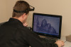
It may be necessary to make wire adjustments (eg, offsets, insets, step-ups, or step-downs) and change specific tubes or brackets that might be off position to enable arches to match correctly. Occlusal adjustment might be needed as well to remove interferences. Presurgical dental models should fit well, and no interference should be present upon simulated surgery movement if analogic planning is used. Surgeons today routinely use virtual planning for highly precise and shorter surgical procedures (Figure 8 and Figure 9).
Surgical Wires
Stainless-steel .018-in x .025-in rectangular wires are steel tied in place a month before surgery in a .022-in slot.1,3,19,26 These wires should have soldered brass wire hooks incorporated 10 to 15 days before surgery. In the authors' practices, these pins in the wires are generally placed in the midlines and anteriorly to the cuspids and first molars in both arches (although, in some instances, more pins might be added as needed).
Surgical wires should be rigid and fill the bracket slot, as the surgeon uses them to lock the occlusion for rigid fixation purposes. The clinician is faced with the choice of commercial pins or tailor-made soldered pins. Soldered hooks are preferred over prefabricated crimpable ones since the former tend not to break when taking presurgical dental impressions. They are also more resistant to forces that may be applied during surgery. Note that as a precaution, when using soldering hooks, it is necessary to avoid excessive heat that might alter the wire rigidity.3
When a posterior segmental osteotomy is planned in cases where premolars will be extracted during surgery to reduce dental protrusion, no pins are placed distal to the sliding segment. In some anterior open bites, the upper arch is segmented into three pieces for a more straightforward surgical procedure. In surgically assisted rapid palatal expansion cases, upper arch surgical wires are not placed so as to avoid interference with transverse expansion. TADs are widely used by surgeons today to complement surgical wires or as a means of surgical fixation in patients with clear aligners because these patients do not have brackets, wires, or surgical pins.
Postsurgical Orthodontics and Finishing the Case
Postsurgically, rigid fixation is fundamental for stability.29 Full recovery after surgery is generally achieved in 3 to 4 weeks. Early physical therapy is a critical step in the authors' postsurgical protocols; it allows for eating solid foods sooner, shorter recovery time, and a quicker return to everyday life for the patient.
The patient is seen 3 to 5 weeks after surgery. The bite interarch settlement is obtained, and the arch coordination is finalized (Figure 10); the occlusion details of the case are finished,30 postsurgical records are taken, and the case is filed.
The literature reports an average of 17 months of presurgical orthodontic preparation.31 In the authors' practices, a full surgical-orthodontic case usually takes between 20 and 22 months, including pre- and postsurgical orthodontics, with the latter usually requiring 4 to 6 months.
In transverse discrepancies corrected with surgical expansion, the Hass, Hyrax, or TAD supported expanders are kept in place for 8 to 10 months to ensure bone deposition in the midline and transverse stability. In two-piece Le Fort I procedures for maxillary expansions, a robust transpalatal .045-in arch from upper first molar to upper first molar is used postsurgery. If simultaneous vertical and transverse correction is sought, multiple-piece Le Fort I osteotomies (more than two pieces) are an option. Surgeons sometimes request a .036-in overlay arch that is placed in the headgear tube. In such cases, these patients require upper first molar bands instead of bonded tubes. Flow resin is added to the upper lateral incisor and cuspid brackets to lock the area where the surgical cut was made. These brackets might need replacement postsurgically. Another consideration may be vertical surgical corrections in open bites, as the six upper anterior brackets might need to be recemented more gingivally postsurgery.
Sequence of Postsurgical Orthodontic Care
The patient returns 1 month after surgery, and surgical wires with the soldered brass pins are removed. Dental cleaning with ultrasound is performed to remove any food particles that were trapped in the surgical wires. The patient is referred to a periodontist if necessary.
Either round stainless-steel .018-in wires or a braided rectangular wire are placed, starting with the use of posterior box elastics to settle the bite and avoiding the use of anterior elastics as much as possible. The posterior elastics might have a vector (class II or class III) if required.30,32
The case is finished using rectangular steel wires for proper tooth positioning. The patient is asked to stop wearing elastics for about 8 weeks (usually the timespan of two appointments) before the orthodontic appliances are removed to determine if the occlusion is stable. The arches are consolidated with a figure-eight ligature from second premolar to second premolar to avoid spaces reopening before debonding.
Before the orthodontic appliances are removed, the occlusion is adjusted with articulating paper once the marginal ridges are well aligned. The orthodontic appliances are then removed and the patient is provided with a retainer.
In summary, the presurgical preparation usually takes about 65% of treatment time. After the surgical repositioning of the bones, the finalization of the postsurgical interdigitation and detailing ensues for a few more months.
Early Vs Late Surgery
If a case meets all preliminary requirements-minimal crowding, good incisal inclination, good transverse arch coordination, healthy TMJs, and absence of genetic syndromes or multiple missing teeth-the patient may possibly be treated with a "surgery first" approach.33 This approach alters the usual sequence of surgical-orthodontic therapy and accounts for shorter treatment time. Often in these cases, early miniplate placement is used for simpler postsurgical movements. Along this line of thinking, some clinicians prefer to perform most of the dental corrections postsurgically.30 In the authors' practices, the more conventional surgical approach is typically used rather than the early surgical approach, and most of the preparation is done prior to surgery.
Re-evaluation of Surgical-Orthodontic Results
Seventy-five percent of surgical cases are very stable.34 Relapse, if present, might be of dental or skeletal origin. Dental relapse is usually limited to minor crowding or spacing. Skeletal relapse may occur during the first year. Upward movement of the maxilla and mandibular surgical advancement up to 9 mm is the most stable surgical procedure. Moderate maxillary advancements of less than 8 mm are next in terms of stability. Moving the maxilla down, setting the mandible back, and performing multiple-piece Le Fort I expansions are the procedures that show more tendency to relapse.34 Adequate planning and a controlled surgical procedure reduce relapse. Virtual guidance accounts for better predictability.
Conclusion
A thorough, accurate diagnosis is key in determining which patients may benefit from a combination of orthodontics and orthognathic surgery. Along with the surgical procedure itself, presurgical and postsurgical orthodontics can be crucial elements in the management of dentofacial deformities. Planning requires managing the technical details of each dental specialty and excellent communication between the orthodontist and oral and maxillofacial surgeon. Critical review of results from facial and occlusal points of view is essential for delivering optimal quality of care.
About the Authors
Miguel Hirschhaut, DDS
Private orthodontic practice, Caracas, Venezuela
Carlos Flores-Mir, DDS, DSc
Professor, Division of Orthodontics, Department of Dentistry, University of Alberta, Edmonton, Alberta, Canada; Part-time Private Practice limited to Orthodontics, Edmonton, Alberta, Canada
Queries to the author regarding this course may be submitted to authorqueries@aegiscomm.com.
References
1. Proffit WR, White RP Jr. Combining surgery and orthodontics: Who does what, when? In: Proffit WR, White RP Jr, Sarver DM, eds. Contemporary Treatment of Dentofacial Deformity. St Louis, MO: Mosby; 2003:245-267.
2. Williams AC, Shah H, Sandy JR, Travess HC. Patients' motivations for treatment and their experiences of orthodontic preparation for orthognathic surgery. J Orthod. 2005;32(3):191-202.
3. Sabri R. Orthodontic objectives in orthognathic surgery: state of the art today. World J Orthod. 2006;7(2):177-191.
4. Betts NJ, Vanarsdall RL, Barber HD, et al. Diagnosis and treatment of transverse maxillary deficiency. Int J Adult Orthodon Orthognath Surg. 1995;10(2):75-96.
5. Vanarsdall RL Jr. Transverse dimension and long-term stability. Semin Orthod. 1999;5(3):171-180.
6. Thiesen G, Gribel BF, Freitas MP. Facial asymmetry: a current review. Dental Press J Orthod. 2015;20(6):110-125.
7. Ricketts RM. Introducing Computerized Cephalometrics. Denver, CO: Rocky Mountain Data Systems, Inc.; 1969.
8. Tamburrino RK, Boucher NS, Vanarsdall RL, Secchi AG. The transverse dimension: diagnosis and relevance to functional occlusion. RWISO Journal. September 2010:13-21.
9. Dym H, Pierre R 2nd. Diagnosis and treatment approaches to a "gummy smile." Dent Clin North Am. 2020;64(2):341-349.
10. Downs WB. Analysis of the dentofacial profile. Angle Orthod. 1956;26(4):191-212.
11. Ricketts RM. Divine proportion in facial esthetics. Clin Plast Surg. 1982;9(4):401-422.
12. Sperry TP, Worms FW, Isaacson RJ, Speidel TM. Tooth-size discrepancy in mandibular prognathism. Am J Orthod. 1977;72(2):183-190.
13. Halimi A, Zaoui F. Surgical-orthodontic treatment of patients suffering from severe periodontal disorders - a clinical case study. Int Orthod. 2013;11(3):314-332.
14. Rozzi M, Mucedero M, Pezzuto C, Cozza P. Leveling the curve of Spee with continuous archwire appliances in different vertical skeletal patterns: a retrospective study. Am J Orthod Dentofacial Orthop. 2017;151(4):758-766.
15. Jiménez-Silva A, Tobar-Reyes J, Vivanco-Coke S, et al. Centric relation-intercuspal position discrepancy and its relationship with temporomandibular disorders. A systematic review. Acta Odontol Scand. 2017;75(7):463-474.
16. Arad I, Jandu J, Bassett P, Fleming PS. Influence of single-jaw surgery vs bimaxillary surgery on the outcome and duration of combined orthodontic-surgical treatment. Angle Orthod. 2011;81(6):983-987.
17. Proffit WR, White RP. The search for truth: diagnosis. In: Proffit WR, White RP, eds. Surgical-Orthodontic Treatment. St. Louis, MO: Mosby; 1991:96-141.
18. Sarver D, Proffit WR, Ackerman JL. Diagnosis and treatment planning in orthodontics. In: Graber TM, Vanarsdall RL, eds. Orthodontics: Current Principles and Techniques. 3rd ed. St Louis, MO: Mosby; 2000:3-115.
19. Larson BE. Orthodontic preparation for orthognathic surgery. Oral Maxillofac Surg Clin North Am. 2014;26(4):441-458.
20. Nocini PF, Chiarini L, Bertossi D. Cosmetic procedures in orthognathic surgery. J Oral Maxillofac Surg. 2011;69(3):716-723.
21. Squire D, Best AM, Lindauer SJ, Laskin DM. Determining the limits of orthodontic treatment of overbite, overjet, and transverse discrepancy: a pilot study. Am J Orthod Dentofacial Orthop. 2006;129(6):804-808.
22. Proffit WR, Phillips C, Tulloch JF, Medland PH. Surgical versus orthodontic correction of skeletal Class II malocclusion in adolescents: effects and indications. Int J Adult Orthodon Orthognath Surg. 1992;7(4):209-220.
23. Hirschhaut M, Flores-Mir C. Orthodontic learning curve - a journey we all go through. Am J Orthod Dentofacial Orthop. In press.
24. Wolford LM. Idiopathic condylar resorption of the temporomandibular joint in teenage girls (cheerleaders syndrome). Proc (Bayl Univ Med Cent). 2001;14(3):246-252.
25. Farret MM. Occlusal plane canting: a treatment alternative using skeletal anchorage. Dental Press J Orthod. 2019;24(1):88-105.
26. Vanarsdall RL Jr. Presurgical orthodontics for orthognathic surgery. Atlas Oral Maxillofac Surg Clin North Am. 2001;9(1):75-93.
27. de Araújo TM, Caldas LD. Tooth extractions in orthodontics: first or second premolars? Dental Press J Orthod. 2019;24(3):88-98.
28. Pandis N, Strigou S, Eliades T. Maxillary incisor torque with conventional and self-ligating brackets: a prospective clinical trial. Orthod Craniofac Res. 2006;9(4):193-198.
29. Van Sickels JE, Richardson DA. Stability of orthognathic surgery: a review of rigid fixation. Br J Oral Maxillofac Surg. 1996;34(4):279-285.
30. Lee RT. The benefits of post-surgical orthodontic treatment. Br J Orthod. 1994;21(3):265-274.
31. Luther F, Morris DO, Hart C. Orthodontic preparation for orthognathic surgery: how long does it take and why? A retrospective study. Br J Oral Maxillofac Surg. 2003;41(6):401-406.
32. Noffel SE. Mandibular incisor uprighting identification. Am J Orthod Dentofacial Orthop. 1995;107(4):426-433.
33. Uribe FA, Farrell B. Surgery-first approach in the orthognathic patient. Oral Maxillofac Surg Clin North Am. 2020;32(1):89-103.
34. Bailey LJ, Cevidanes LH, Proffit WR. Stability and predictability of orthognathic surgery. Am J Orthod Dentofacial Orthop. 2004;126(3):273-277.