You must be signed in to read the rest of this article.
Registration on AEGIS Dental Network is free. Sign up today!
Forgot your password? Click Here!
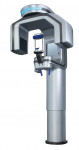
PreXion3D Excelsior Visualizing anatomy in 3D improves diagnosis for all clinical applications
Jon Julian, DDS
Private Practice Travelers Rest,
South Carolina
In 40 years of dentistry, the most essential thing I have learned is the importance of accurately diagnosing my patients' conditions. To this end, I became a user of cone-beam computed tomography (CBCT) 10 years ago when I purchased my first unit from PreXion.
When I started seeing hard-tissue structures in 3 dimensions, a world of discovery was opened to me. Although my previous 2-dimensional radiographs often left me guessing when diagnosing a patient's problem, with the CBCT images obtained, my patients and I were able to clearly see the existing problems. As I gained experience, I found that I was taking CBCT images for most of my patients, primarily to screen them for existing problems or to reassure them that no pathologies were present. Oftentimes, I found real issues that the patient was unaware of. When patients are shown their conditions and provided with education, they accept recommendations more readily and are able to better understand and participate in planning their treatment.
Furthermore, the CBCT scan-ner revealed periapical lesions that were not visible on my 2D films. Their size and location were unmistakable. I could also accurately visualize the position of all third molars and their relationship to vital structures, which helped me in my decisions to treat or refer these cases. Because I could see the number of canals as well as their shape and length prior to treatment, my decisions to treat or refer endodontic cases were better informed, as well. Prior to beginning any restorative treatment, I could see the extent of caries and their proximity to the pulp chamber. My periodontal therapy also improved because I could visualize defects in 3D, especially the furcations of maxillary molars. And finally, the patient's airway is visible on every scan, which helps open the door to anyone interested in sleep dentistry. I can do all of the aforementioned and more while exposing my patients to less than half of the radiation that they would be exposed to during a full-mouth series of of 2D films. Patients really appreciate this aspect of the scanner.
The PreXion3D Excelsior delivers superior image quality because it has a small focal spot size, a small voxel size, 512 image slices, and a quick scan capture time. From a doctor's perspective, it has very intuitive software that is easy to learn and features free upgrades. In addition, the technical support is world class, and education beyond the initial training is included, which allows the doctor to gain additional experience over time.
If this resonates with you, and you are in the market for a CBCT scanner, compare the PreXion3D Excelsior side-by-side with any other scanner on the market, and you'll see why it came out on top for me.
Key Takeaways
1. Accurate 360° gantry rotation with 260 to 1,024 projected views
2. Includes PreXion3D Viewer software at no additional charge
3. Can be used for various clinical applications, including implant placement, endodontics, airway analysis, and more
4. Provides clinicians with themost accurate assessment of the bone and surrounding anatomy while making exact 1:1 measurements
Manufacturer Information
PreXion
prexion.com
855-773-9466
Circle 61 on Reader Service Card