You must be signed in to read the rest of this article.
Registration on CDEWorld is free. You may also login to CDEWorld with your DentalAegis.com account.
The predictability of any implant restoration greatly depends on the treatment team adopting a restoratively driven, or "end-in-mind," approach in treatment planning. Understanding and visualizing the final prosthesis before initiating surgical treatment is key to achieving an anticipated outcome.
Evaluation of edentulous patients, especially those with edentulous maxillae, can be complicated due to the fact that patients may only be missing clinical crowns, ie, a tooth-only defect, or they may present with a combination of tooth loss and soft- and hard-tissue loss, ie, a composite defect.1 Patients with terminal dentition typically present with bone and soft-tissue loss having occurred before tooth removal as a result of generalized periodontitis, creating the appearance of long teeth; in the authors' experience these types of patients usually present with a composite defect, too. Thus, a systematic treatment planning protocol would be beneficial for the evaluation of both edentulous patients and those who present with terminal dentition.
The loss of teeth and use of a removable prosthesis can result in continued alveolar bone atrophy in both vertical and horizontal dimensions. In a study spanning 25 years, Tallgren observed that the greatest amount of alveolar bone atrophy occurs within the first year of edentulism.2 Also, changes in the jaw relationship and in facial musculature may result in deformation or other changes in the facial form and morphology.3 A systematic pretreatment approach allows for improved communication among the implant team and with the patients, which typically helps in achieving a predictable treatment outcome.
Three important factors that can be determined early in the examination process can be key to the successful treatment of the completely edentulous maxilla with a fixed restoration. These factors are: (1) the presence or absence of a composite defect, which helps determine the type of final prosthesis; (2) visibility of the transition line, which is used for evaluation for an esthetic outcome; and (3) the available alveolar bone in the edentulous maxilla, which determines the surgical protocol. While not intended to be a substitute for thorough diagnosis and development of a treatment plan, evaluation of these three factors can provide differential diagnosis information specific to the esthetic, phonetic, and biomechanical requirements of fixed, implant-supported maxillary restorations.
Presence or Absence of a Composite Defect
The clinical decision-making algorithm for ascertaining the type of final prosthesis appropriate for a patient is impossible to determine without first identifying whether a patient has a tooth-only defect or a composite defect. Edentulous patients may present with intact alveolar bone volume and missing only clinical crowns (tooth-only defect), or they may present with alveolar bone resorption, loss of soft tissue, and missing teeth (composite defect). Differentiating between these two types of patients is crucial to creating an esthetic definitive fixed prosthesis.
Patients who present with missing teeth without any resorption of the soft or hard tissues are referred to as having a "tooth-only defect." To confirm that a tooth-only defect is present, the clinician can examine the space between the edentulous crest and the cervical portion of clear denture teeth replicated from the patient's existing denture. The presence of no space as seen through the clear denture, ie, the cervical portion of the teeth resting on crestal soft tissues, confirms the diagnosis of a tooth-only defect. For this group of patients, a fixed "white" ceramo-metal or all-ceramic restoration may be planned.
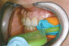
The use of a clear denture also can aid in establishing the diagnosis of a "composite defect," which is the term describing patients who are missing teeth as well as soft and hard tissues. The presence of space between the edentulous crest and the cervical portion of the clear denture teeth confirms the presence of a composite defect (Figure 1). For this group of patients, a fixed hybrid prosthesis or an implant-supported overdenture may be planned.
For the clinician to confidently evaluate the relative amount of soft- and hard-tissue deficiency, the clear denture duplicated from the patient's existing denture must have the correct vertical dimension of occlusion as well as the proper anterior-posterior and horizontal tooth position.
Visibility of Residual Ridge Crest: the Transition Line
Once the presence or absence of a composite defect has been established, the transition line must be evaluated for patients for whom a maxillary hybrid prosthesis is planned. If a composite defect is present, it would be inappropriate to plan a metal-ceramic or all-ceramic tooth-only restoration because such a restoration would result in esthetic compromises due to longer-than-normal teeth. Therefore, a hybrid prosthesis, ie, a "pink and white" prosthesis, intended to replace the teeth and missing hard and soft tissues should be planned. To maximize the esthetic prosthetic outcome for this group of patients, the transition line between the hybrid prosthesis and the soft tissue of the edentulous maxillary ridge must be clinically evaluated for potential visibility without the maxillary denture in place.
This evaluation can be done by removing the maxillary denture and then having the patient smile (Figure 2). If the soft tissue of the edentulous ridge cannot be seen, the transition between an implant-supported hybrid prosthesis and the residual soft-tissue crest is considered favorable. Conversely, for those patients who do display the residual ridge while smiling, ie, the smile line is apical to the transition line, the transition between a hybrid restoration and the soft tissue will be visible and, therefore, unesthetic. With a visible residual ridge crest, the junction of the artificial gingiva of the hybrid prosthesis and the natural soft tissue will be apparent, and differences in texture and contour between the two may be obvious and unesthetic. However, if the transition line is apical to the smile line, ie, the smile line is incisal to the transition line, a predictable esthetic outcome is possible.
If the crestal soft tissues are visible in the preoperative evaluation, one method to avoid visibility of the transition line in the final hybrid prosthesis is to reduce the residual ridge height to the point where the crest can no longer be seen and is apical to the smile line prior to placement of implants. Thus, an intentional alveoloplasty in conjunction with implant placement would be planned. If ridge reduction is not possible due to large pneumatized maxillary sinuses, the use of a Marius bridge or any variation of an implant-supported overdenture with a flange that overlaps the gingival junction would be indicated.
In contrast, for patients who are missing teeth only, the visibility or lack of visibility of the transition line is not an issue, because the implants are placed in planned tooth positions, and special consideration is given to anterior ridge lap pontics to improve the appearance of papillae and achieve an esthetic outcome (Figure 3). The emergence profile of the prosthesis from the implant platform should be such that the resultant fixed "white" bridge mimics the emergence profile of the patient's teeth prior to their extraction.
Radiographic Evaluation
Radiographic evaluation of the edentulous maxilla is necessary for determining whether axial, tilted, or zygomatic implants would be indicated to establish optimal posterior support with proper anterior-posterior distribution of implants for a fixed prosthesis. Because the edentulous maxilla is divided into three radiographic zones, a systematic assessment of the residual alveolar bone available for implant placement can be made. In this pretreatment screening protocol, the alveolar bone supporting the maxillary anterior teeth is designated as zone 1, while the premolar region is considered zone 2 and the molar region zone 3 (Figure 4). Analysis of the radiographic results according to this scheme can enable the surgical and restorative team to devise a preliminary treatment plan. In complex situations, 3-dimensional (3D) radiographic evaluation may still be necessary to confirm the preliminary conclusions.
For fully fixed implant-supported maxillary restorations, the distribution of the implants along the arch form is as important as the number of implants used. The goal is to place the posterior implants as far posterior as possible from the anterior implants to achieve the largest anterior-posterior distribution of the implants as possible. A minimum of four implants should be used, although the option to place more than four may be considered.4,5 As a general principle, cantilevers in fixed maxillary restorations should be avoided or minimized to one tooth to avoid unfavorable load transfer to the prosthetic components, alveolar bone, and existing implants.6,7
Evaluation of the various zones of the maxilla will guide the surgical team in determining the type of surgical approach to take for placement of implants. Use of panoramic scout film as well as 3D radiography to evaluate the zones will help the surgeon understand the quantity of bone available for each intended implant site.
Presence of Zone 1, 2, and 3 Bone
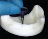
For patients in whom alveolar bone is present in all three zones of the edentulous maxilla, conventional implants may be placed (Figure 5). This should allow for a favorable arch form of anterior, posterior, and possibly intermediate implants for a fixed prosthesis.8,9
Presence of Zone 1 and 2 Bone
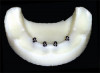
For patients who have zone 1 and zone 2 bone but lack zone 3 bone secondary to large pneumatized maxillary sinuses, inclining the implants posteriorly along the anterior wall of the maxillary sinus may enable an adequate anterior and posterior distribution of implants to support a fixed restoration while avoiding the need for grafting (Figure 6).4,10-15 Use of inclined implants also has been shown to be successful with immediate-loading procedures of the completely edentulous maxilla.5,13,15
An alternative to the use of inclined implants is sinus inlay grafting, followed by subsequent delayed implant placement. When extensive sinus inlay grafting is performed to provide posterior support, a staged approach that incorporates an allotment of time to wait for graft maturation may be preferable due to lower survival for implants that are simultaneously placed and loaded.16 This method has the effect of delaying restoration compared with the use of inclined implants and a graftless approach.
Presence of Zone 1 Bone Only
To establish posterior support for a fixed prosthesis, implants are required in the second premolar or first molar regions. However, placement of implants in these positions is not possible when patients only have bone available in zone 1. Grafting of the sinus with autogenous bone or xenografts is an option in such situations. This approach has shown a 90% overall survival rate with 3- to 5-year follow-up.17
If a graftless approach is pre-ferred, zygomatic implants have been shown to provide bilateral posterior maxillary support with a 97% to 100% survival rate.18-20 These implants have the added benefit of not requiring a staged approach or a period of bone graft maturation. Thus, the overall treatment time required to achieve a fixed restoration can be shortened. Predictable posterior support can be established with the placement of one zygomatic implant in each zygoma.19,20 When zygomatic implants are used in conjunction with two to four axial anterior implants, a fixed, implant-supported prosthesis may be fabricated (Figure 7).
Bone Missing From Zones 1, 2, and 3
When there is complete resorption of the maxillary alveolus, clinical examination will reveal a flat palatal vault. No maxillary vestibule will be present, and the patient typically is unable to function with his or her conventional complete denture. In the authors' experience such patients usually present with a significantly thick denture base and a thick circumferential flange, confirming the presence of a significant composite defect.
Physiologic reconstruction of these debilitated patients requires adequate implant support to stabilize an implant-supported prosthesis. To enable prosthetic rehabilitation of such patients, Bränemark introduced the idea of using extensive onlay bone grafts in conjunction with bilateral sinus inlay grafts and placement of six implants.21 The so-called Brånemark "horseshoe" graft requires hospitalization and harvesting of autogenous iliac bone from the patient (Figure 8). These patients are unable to wear a denture during the 6-month osseointegration period. The social consequence of this form of treatment renders it unpopular with patients.
An alternative, graftless approach is the use of four zygomatic implants (Figure 9). The placement of two such implants in each zygoma allows for the fabrication of an implant-supported fixed maxillary prosthesis without the need for bone grafting and can be accomplished in an office setting. In 2015 Wang et al reported a 96.7% success rate in a systematic review of quad-zygoma implants supporting fixed maxillary hybrid prostheses.22
Discussion
Many factors must be considered before implant treatment is performed in a patient with a fully edentulous maxilla. Pretreatment screening can be used to identify early whether or not it is likely that the patient's expectations will be satisfied with a prosthesis option that realistically takes into account not only tooth loss but also the amount of soft- and hard-tissue deficit that must be restored. Additionally, systematic panoramic and 3D radiograph analysis based on available zones of the edentulous maxilla can provide an early indication of how straightforward or difficult the surgical treatment might be.
The combination of prosthodontic and radiographic diagnostic criteria can offer an early indication of treatment possibilities from both surgical and restorative perspectives to help the dental team clarify and communicate the potential treatment requirements and desired outcome. The dental team may then use this information to suggest to the patient to proceed with further, more definitive diagnostics, confident that the anticipated prosthetic outcome may be possible. However, it must be noted that the critical factor of sufficient alveolar ridge width still needs to be verified, and this would only be discovered after a scan. Lack of sufficient ridge width could change the surgical approach significantly.
Clinicians should also bear in mind that these diagnostic criteria still need to be evaluated in relation to the patient's overall health and medical and dental history. Additionally, it must be clearly understood that even with the most thorough planning, deviations from the desired outcome may occur. The criteria presented in this article are best viewed as a preliminary screening apparatus to help guide patient and clinical decisions as more information is gathered. They are subject to change at any time if more definitive analysis or radiographic information does not support the preliminary indication. Also, there may be clinical situations where the objective is to remove remaining hopeless teeth and simultaneously place implants. While this preliminary diagnostic method is still applicable in these situations, it cannot account for variations in tissue height that may result subsequent to dental extraction.
Conclusion
This article discussed a pretreatment screening method that systematically considers the presence or absence of a composite defect, the visibility of the residual soft-tissue crest, and the availability of bone in three radiographic zones as guidelines for the selection of three potential fixed implant restorative designs: the "all-white" metal-ceramic or all-ceramic prosthesis, the fixed hybrid prosthesis, or the implant-supported overdenture. Additionally, this screening method offers guidance on the optimal implant surgical approach, including the use of axial, tilted, and/or zygomatic implants. By employing these differential diagnosis criteria the dental team is able to make an early determination of the treatment necessary to meet patient expectations before investing a significant amount of time and resources.
A limitation of this protocol is the inability to measure the width of the available residual alveolar bone. While panoramic survey film provides a valuable 2-dimensional scouting radiograph and allows the practitioner to evaluate the height and length of the residual alveolar bone, 3D tomography or spiral computed tomography studies can be used to precisely measure the width of the remaining ridge to aid in making a final determination of the likely outcome of the planned treatment. Adoption of this evaluation method may improve the uniformity of communication among dental colleagues, laboratory support, third-party payment providers, as well as students and faculty.
About the Authors
Edmond Bedrossian, DDS
Professor, Department of Oral & Maxillofacial Surgery, Arthur A. Dugoni School of Dentistry, University of the Pacific, San Francisco, California; Diplomate, American Board of Oral & Maxillofacial Surgery; Honorary Member, American College of Prosthodontists
Edmond Armand Bedrossian, DDS
Private Practice, Prosthodontist, San Francisco, California
Queries to the author regarding this course may be submitted to authorqueries@aegiscomm.com.
References
1. Bedrossian E, Sullivan RM, Fortin Y, et al. Fixed-prosthetic implant restoration of the edentulous maxilla: a systematic pretreatment evaluation method. J Oral Maxillofac Surg. 2008;66(1):112-122.
2. Tallgren A. The continuing reduction of the residual alveolar ridges in complete denture wearers: a mixed-longitudinal study covering 25 years. J Prosthet Dent. 2003;89(5):427-435.
3. Cawood JI, Howell RA. Reconstructive preprosthetic surgery. I. Anatomical considerations. Int J Oral Maxillofac Surg. 1991;20(2):75-82.
4. Brånemark PI, Svensson B, van Steenberghe D. Ten-year survival rates of fixed prostheses on four or six implants ad modum Brånemark in full edentulism. Clin Oral Implants Res. 1995;6(4):227-231.
5. Maló P, Rangert B, Nobre M. All-on-4 immediate-function concept with Brånemark System implants for completely edentulous maxillae: a 1-year retrospective clinical study. Clin Implant Dent Relat Res. 2005;7 suppl 1:S88-S94.
6. Sadowsky SJ. The implant-supported prosthesis for the edentulous arch: design considerations. J Prosthet Dent. 1997;78(1):28-33.
7. Taylor TD. Fixed implant rehabilitation for the edentulous maxilla. Int J Oral Maxillofac Implants. 1991;6(3):329-337.
8. Renouard F, Rangert B. Risk Factors in Implant Dentistry. Chicago, IL: Quintessence Publishing; 1999:103-109.
9. Engelman MJ. Clinical Decision Making and Treatment Planning in Osseointegration. Chicago, IL: Quintessence Publishing; 1996.
10. Jemt T. Failures and complications in 391 consecutively inserted fixed prostheses supported by Brånemark implants in edentulous jaws: a study of treatment from the time of prosthesis placement to the first annual checkup. Int J Oral Maxillofac Implants. 1991;6(3):270-276.
11. Mattsson T, Köndell P, Gynther GW, et al. Implant treatment without bone grafting in severely resorbed edentulous maxillae. J Oral Maxillofac Surg. 1999;57(3):281-287.
12. Krekmanov L, Kahn M, Rangert B, Lindström H. Tilting of posterior mandibular and maxillary implants for improved prosthesis support. Int J Oral Maxillofac Implants. 2000;15(3):405-414.
13. Krekmanov L. Placement of posterior mandibular and maxillary implants in patients with severe bone deficiency: a clinical report of procedure. Int J Oral Maxillofac Implants. 2000;15(5):722-730.
14. Aparicio C, Perales P, Rangert B. Tilted implants as an alternative to maxillary sinus grafting: a clinical, radiologic, and periotest study. Clin Implant Dent Relat Res. 2001;3(1):39-49.
15. Calandriello R, Tomatis M. Simplified treatment of the atrophic posterior maxilla via immediate/early function and tilted implants: a prospective 1-year clinical study. Clin Implant Dent Relat Res. 2005;7 suppl 1:S1-S12.
16. Kahnberg KE, Ekestubbe A, Gröndahl K, et al. Sinus lifting procedure. I. One-stage surgery with bone transplant and implants. Clin Oral Implants Res. 2001;12(5):479-487.
17. Jensen OT, Shulman LB, Block MS, Iacono VJ. Report of the Sinus Consensus Conference of 1996. Int J Oral Maxillofac Implants. 1998;13 suppl:11-45.
18. Bedrossian E, Stumpel L III, Beckely ML, Indresano T. The zygomatic implant: preliminary data on treatment of severely resorbed maxillae. A clinical report. Int J Oral Maxillofac Implants. 2002;17(6):861-865.
19. Bedrossian E, Stumpel LJ III. Immediate stabilization at stage II of zygomatic implants: rationale and technique. J Prosthet Dent. 2001;86
(1):10-14.
20. Malevez C, Abarca M, Durdu F, Daelemans P. Clinical outcome of 103 consecutive zygomatic implants: a 6-48 months follow-up study. Clin Oral Implants Res. 2004;15(1):18-22.
21. Breine U, Brånemark PI. Reconstruction of alveolar jaw bone. An experimental and clinical study of immediate and preformed autologous bone grafts in combination with osseointegrated implants. Scand J Plast Reconstr Surg. 1980;14(1):23-48.
22. Wang F, Monje A, Lin GH, et al. Reliability of four zygomatic implant-supported prostheses for the rehabilitation of the atrophic maxilla: a systematic review. Int J Oral Maxillofac Implants. 2015;30(2):293-298.